


Multiple studies have corroborated the association between bullous pemphigoid (BP) and neurological diseases; patients with both diseases (BP-N) have been associated with a worse prognosis and specific clinical and immunological characteristics, defining a different subtype of BP.
ObjectivesWe aimed to determine the prevalence and characteristics of BP cases with neurological comorbidities (BP-N) and review the related published literature.
MethodsWe conducted a retrospective, observational study of BP cases treated at a referral center for autoimmune blistering diseases from January 2000 through June 2020.
ResultsWe collected epidemiological, clinical, histopathological, progression and laboratory data from a total of 257 cases, 102 of which were BP-N. Senile dementia, Alzheimer's disease and vascular dementia were the most frequent neurological comorbidities. Compared with cases without neurological comorbidities, BP-N cases had more intense tissue eosinophilia (p=0.012) and higher concentrations of circulating eosinophils (p=0.000), and anti-BP180 IgG antibodies (p=0.007). At the time of data collection, 78 BP-N were deceased.
ConclusionsOur case series highlighted the relevance of neurological comorbidities in BP patients; although the pathogenesis is still to be elucidated, the neuroinflammation present in neurodegenerative diseases could explain the neurocutaneous link and the chronological relationship between these entities.
Múltiples estudios han corroborado la asociación entre penfigoide ampolloso (PA) y enfermedad neurológica; en ellos, se ha planteado que los pacientes con PA asociado a enfermedad neurológica tendrían unas características clínicas e inmunológicas específicas, condicionando un subtipo especial de PA, que contaría con un peor pronóstico.
ObjetivosDeterminar la prevalencia y características de los casos de PA asociados a comorbilidades neurológicas (PA-N) y revisar la literatura publicada al respecto hasta la fecha.
MétodosEstudio retrospectivo observacional de los casos de PA atendidos entre enero del 2000 y junio del 2020 en una consulta monográfica de enfermedades ampollosas autoinmunes.
ResultadosSe determinaron las características epidemiológicas, clínicas, histopatológicas, inmunológicas y evolutivas de 257 pacientes, de los cuales 102 presentaban comorbilidad neurológica. La demencia senil, enfermedad de Alzheimer y la demencia vascular fueron las más frecuentes. En comparación con pacientes con PA sin enfermedad neurológica, los casos con esta comorbilidad presentaron mayores concentraciones de eosinófilos en sangre (p=0,000) y de anticuerpos anti-BP180 IgG (p=0,007) además de una mayor eosinofilia tisular (p=0,012). En el momento de la recogida de datos, 78 pacientes con PA-N se encontraban fallecidos.
ConclusionesEste estudio refleja la relevancia de las enfermedades neurológicas en pacientes con PA. Aunque la fisiopatogenia de esta asociación no ha sido bien establecida, la neuroinflamación presente en las enfermedades neurológicas, especialmente degenerativas, podría explicar la conexión neurocutánea y la relación cronológica entre ambas enfermedades.

Bullous pemphigoid (BP) is the most common autoimmune blistering disease of the skin and mucous membranes.1–3 Although defined as a rare condition, the incidence rate of BP has been increasing throughout time, attributed to factors such as population aging, neurological and neoplastic comorbidities, drug-induced cases and the increasing awareness of non-bullous and atypical variants of BP.1–4
BP predominantly affects patients older than 65 years and the risk of developing it increases with age.2,3 In a typical patient, BP has been classically associated with neurological conditions,5,6 which are closely associated with advanced age3,5 and have shown an increasing prevalence in the general population.7 Moreover, drug-related BP cases have contributed to the rising incidence rate.8,9 Psychotropic and central nervous system drugs have been identified as potential inducers of dermatological conditions, including BP.4,8,9
Several studies have characterized cases of BP with neurological comorbidities (BP-N)10–16 showing specific clinical and serological characteristics,11 worse prognosis12 and higher mortality rates.13 However, most of the studies were conducted in small samples and focused on epidemiological aspects, often failing to include a control group (BP patients without neurological diseases).12–14
Therefore, the objectives of this study were to determine the prevalence BP-N, including all BP patients treated at a tertiary referral center for autoimmune blistering diseases, and evaluate the clinicopathological and immunological features, management, progression and prognosis of BP-N cases vs cases of BP without neurological comorbidities.
MethodsWe conducted a retrospective, observational study to analyze the relationship between BP and neurodegeneration in patients treated from January 2000 through June 2020.
The diagnosis of BP was based on the criteria proposed by the European Dermatology Forum in collaboration with the European Academy of Dermatology and Venereology, as updated in their clinical practice guidelines on the management of BP.17 Only confirmed cases meeting, at least, 3 out of the 4 criteria (clinical, histopathological, serological [including indirect immunofluorescence (IIF) and/or enzyme-linked immunosorbent assay (ELISA)] and direct immunofluorescence (DIF)) were included. Patients with other dermatological conditions different from BP were excluded.
Data were collected from the databases of the dermatology and dermatopathology departments including age at onset; sex; date and comorbidities at BP diagnosis; drug use within 6 months prior to diagnosis of BP; clinical characteristics, including body surface area (BSA) affected, location, presence of pruritus, symmetry and scalp and mucous membranes involvement; absolute eosinophil count at diagnosis; serum antibody profile (IIF microscopy and ELISA); histopathological findings (subepidermal blistering, inflammatory infiltrate and eosinophilic infiltrate intensity); DIF pattern; treatment for BP; need for hospitalization; and cause and date of death, particularly in BP-N.
Neurological morbidities were assessed based on whether they preceded the onset of BP or were developed during its course, as precise dates of diagnosis and medication were difficult to ascertain based, only, on health records.
BP cases were categorized into 4 groups based on BSA involvement: generalized (>50%), trunk and extremities (<50%), trunk (<50%), extremities (<50%) and other (when not corresponding to prior criteria). Scalp and mucosal involvement were also recorded.
Due to lack of information drawn from the health records, we were unable to assess the presence of an inflammatory/non-inflammatory phenotype or estimate any disease severity score (BPDAI or IGA).
Skin biopsy with hematoxylin and eosin (H&E) stain and DIF was used in all patients included in the study. Skin lesion samples were reviewed to evaluate the presence of subepidermal bullae and inflammatory infiltrate and the intensity of the eosinophilic infiltrate.
In relation to the eosinophilic infiltrate, 6μm sections from skin lesion samples were stained with hematoxylin–eosin (H&E) to evaluate intensity. Representative hotspots were identified and reviewed by 2 different expert dermatopathologists and 2 dermatologists; the number of eosinophils was assessed using a 40× high power field (HPF) objective (scale bar: 100μm). We distinguished a total of 3 categories: present, very intense (++), when >21; present, not very intense (+), when 5–20; not present or scant (−), when <5eosinophils/HPF.
To assess the level of the blister and differentiate BP from other subepidermal bullous diseases, type IV collagen immunohistochemical staining was used. Staining at the dermal portion of the bullae was considered to be compatible.
Anti-BP180 IgG antibodies detected were the ones directed vs the noncollagenous 16A domain (NC16A). This laboratory test was included in a commercially available ELISA kit that included the detection of IgG autoantibodies vs desmoglein 1, desmoglein 3, BP180 and type VII collagen (“MESACUP anti-Skin profile TEST”). Full-length BP180 and BP230 autoantibodies detection were not tested due to unavailability. Anti-epidermal basement membrane and anti-intercellular cement substance antibodies were detected using IIF.
Statistical analysisCategorical variables were expressed as total number with percentages and the continuous ones as means, with standard deviation in symmetric distributions or medians and interquartile ranges in asymmetric variables.
Demographic, clinical, histopathological and laboratory characteristics were compared between BP-N and non-BP-N groups, using the chi-square test (or Fisher's exact test when necessary) for categorical variables and the Student's t-test (or Mann–Whitney U-test/Wilcoxon W-test when necessary) for continuous variables. Bilateral f-test was used for the equal variance test. Statistical analysis was performed using Stata (version 16 StataCorp, College Station, Texas, United States).
ResultsA total of 257 BP cases were included, 59.9% of whom were men. The mean age at diagnosis was 80.49±10.83 years. Of these, 102 (39.7%) were diagnosed with a neurological disease at the time of BP diagnosis, with senile dementia, Alzheimer's disease and vascular dementia being the most common ones. In all cases, the neurological condition preceded the onset of BP. No patients were diagnosed with >1 neurological illness. Neurological comorbidities of the patients included in this study are shown in Table 1.
Neurological comorbidities in patients with BP included in the study.
| Neurological disease | BP-N (n, %)(n=102) | All patients with BP (n, %)(n=257) |
|---|---|---|
| Senile dementia | 26 (25.5) | 26 (10.1) |
| Alzheimer's disease | 26 (25.5) | 26 (10.1) |
| Vascular dementia | 26 (25.5) | 26 (10.1) |
| Parkinson's disease | 17 (16.6) | 17 (6.6) |
| Amyotrophic lateral sclerosis | 3 (2.9) | 3 (1.2) |
| Multiple sclerosis | 2 (2) | 2 (0.8) |
| Lewy body dementia | 2 (2) | 2 (0.8) |
BP: bullous pemphigoid; BP-N: bullous pemphigoid patients with neurological comorbidities.
In 60 cases, new drugs were introduced within a matter of 6 months prior to BP diagnosis including psychotropic drugs [donepezil (n=1) and lacosamide (n=1)], antibiotics [ciprofloxacin (n=2), levofloxacin (n=1), moxifloxacin (n=1) and rifampicin (n=1)] and dipeptidyl peptidase 4 inhibitors (DPP4i) [vildagliptin (n=27), linagliptin (n=17), sitagliptin (n=5) and saxagliptin (n=2)]. A total of 19 patients in whom the introduction of DPP4i was present and 2 cases of psychotropic drugs (donepezil and lacosamide) corresponded to the BP-N group.
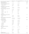
There were no significant differences in the epidemiological and clinical characteristics (BSA) across the groups (Table 2).
Inter-group comparison of the epidemiological and clinical features.
| BP-N (n, %)n=102 | Non-neurological BP (n, %)n=155 | p-Value | |
|---|---|---|---|
| Mean age (±SD) (years) | 81.66 (±9.06) | 77 (±10.88) | 0.078 |
| Sex | 0.771 | ||
| Female | 42 (41.2) | 61 (39.4) | |
| Male | 60 (58.8) | 94 (60.6) | |
| Clinical presentation (% affected BSA) | 0.163 | ||
| Generalized (>50%) | 35 (34.3) | 46 (29.6) | |
| Trunk and extremities (<50%) | 51 (50.0) | 71 (45.8) | |
| Trunk (<50%) | 5 (4.9) | 8 (5.2) | |
| Extremities (<50%) | 11 (10.8) | 28 (18.1) | |
| Other (<50%) | 0 | 2 (1.3) | |
| Scalp involvement | 0.563 | ||
| Yes | 10 (9.8) | 12 (7.7) | |
| No | 92 (90.2) | 143 (92.3) | |
| Mucosal involvement | 0.853 | ||
| Yes | 6 (5.9) | 10 (6.5) | |
| No | 96 (94.1) | 145 (93.5) | |
| Pruritus | 102 (100) | 155 (100) | 0.764 |
| Symmetrical lesions | 0.689 | ||
| Yes | 97 (95.1) | 149 (96.1) | |
| No | 5 (4.9) | 6 (3.9) | |
BP: bullous pemphigoid; BP-N: bullous pemphigoid patients with neurological comorbidities; BSA: body surface area; NS: not significant.
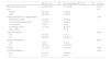
However, immunopathological and serological differences were notable across the groups (Table 3). Despite the absence of differences in blood eosinophilia at diagnosis, the median absolute eosinophil count was higher in the BP-N group (p=0.000).
Immunological, histopathological, progression characteristics, and inter-group comparison.
| BP-N (n, %)n=102 | Non-neurological BP (n, %)n=155 | p-Value | |
|---|---|---|---|
| Blood eosinophilia (>500cells/mm3) | 32 (31.4) | 37 (23.9) | 0.163 |
| Absolute eosinophil count | 0.000 | ||
| Median [IQR] (cells/mm3) | 500 [700] | 300 [575] | |
| Intensity of eosinophilic infiltrate (H&E) | 0.012 | ||
| Present, very intense (++) | 78 (76.5) | 96 (62) | |
| Present, not intense (+) | 22 (21.5) | 57 (36.8) | |
| Not present (−) | 2 (2) | 2 (1.2) | |
| Histopathological characteristics (H&E) | |||
| Subepidermal blistering | 102 (100) | 154 (99.4) | 0.235 |
| Inflammatory infiltrate | 100 (98.3) | 152 (98.6) | 0.633 |
| Direct immunofluorescence pattern (DIF) | 0.173 | ||
| Linear IgG+C3 | 76 (74.5) | 96 (61.9) | |
| Linear C3 | 20 (19.5) | 40 (25.8) | |
| Linear IgG | 2 (2) | 3 (1.9) | |
| Linear IgG+C3+IgM | 2 (2) | 9 (5.8) | |
| Linear C3+IgM | 1 (1) | 0 | |
| Linear C3+IgM+IgA | 1 (1) | 1 (0.6) | |
| Fibrinogen | 0 | 5 (3.2) | |
| Negative DIF | 0 | 1 (0.6) | |
| Anti-BP180 antibodies detection (ELISA) | 0.143 | ||
| Positive (>9U/mL) | 49 (48.3) | 66 (42.9) | |
| Negative | 17 (16.7) | 38 (22.4) | |
| Total | 66 (64.7) | 104 (67.1) | |
| Anti-BP180 antibodies concentration (ELISA) | 0.007 | ||
| Mean (±SD) (U/mL) | 62.06 (±57.8) | 40.24 (±48.9) | |
| Median (IQR) (U/mL) | 47.44 [95.02] | 16.36 [64.1] | |
| Prescribed treatments | 0.035 | ||
| Topical corticosteroids | 102 (100) | 155 (100) | |
| Systemic corticosteroids | 94 (92.2) | 122 (78.7) | |
| <0.5mg/kg/day | 77 (75.5) | 101 (65.2) | |
| >0.5mg/kg/day | 17 (16.7) | 11 (7.1) | |
| Tetraciclines | 12 (11.8) | 17 (10.9) | |
| Azatioprine | 25 (24.5) | 38 (24.5) | |
| Methotrexate | 8 (7.8) | 13 (8.4) | |
| Dapsone | 4 (3.9) | 7 (4.5) | |
| Rituximab | 0 | 6 (3.9) | |
| Number of prescribed treatments | 0.253 | ||
| Mean (±SD) | 1.88 (±0.81) | 2.18 (±0.98) | |
| Median | 2 | 2 | |
| Number of recurrences | 0.181 | ||
| 0 | 83 (81.4) | 115 (74.2) | |
| 1 | 15 (14.7) | 30 (19.4) | |
| 2 | 3 (2.9) | 7 (4.5) | |
| 3 | 1 (1) | 2 (1.3) | |
| 4 | 0 | 1 (0.6) | |
| Hospitalization (days) | 38 (37.5) | 55 (35.5) | 0.773 |
| Deceased at the time of data collection | 78 (76.5) | 77 (49.7) | 0.000 |
BP: bullous pemphigoid; BP-N: bullous pemphigoid patients with neurological comorbidities; C3: C3 complement; DIF: direct immunofluorescence; ELISA: enzyme-linked immunosorbent assay; IgA: immunoglobulin A; IgG: immunoglobulin G; IgM: immunoglobulin M; IQR: interquartile range (p25–p75); H&E: hematoxylin–eosin; mm3: cubic milimeter; n: total number; NS: not significant.
Moreover, compared with the non-neurological group, the BP-N group had elevated eosinophilic inflammatory infiltration (tissular eosinophilia) which was quantified as “present, very intense” (76.5% vs 62%) (p=0.012) (Table 3).
Compared with the non-neurological BP group, BP-N patients had a similar positivity rate (>9U/mL) of anti-BP180-NC16A IgG (p=0.143), but a higher concentration of these antibodies (p=0.007). Serological studies were performed in 66 patients (64.7%) of the BP-N group.
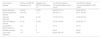
The highest median anti-BP180 antibodies titer was found among patients with Alzheimer's disease [84.01U/mL, interquartile range (IQR) 87.43], followed very closely by multiple sclerosis (86.93U/mL). Vascular dementia exhibited the lowest median antibodies titer (12.23U/mL, IQR 61.26) (Table 4).
Rate of detection and concentration of antiBP-180 autoantibodies based on neurological comorbidities.
| Neurological disease | Positive anti-BP180 antibodies(n, %) | Negative anti-BP180 antibodies(n, %) | Anti-BP180 antibody concentrationMean (±DS) (U/mL) | Anti-BP180 antibody concentrationMedian (IQR) (U/mL) |
|---|---|---|---|---|
| Senile dementia | 10 (9.8) | 3 (2.9) | 54.23 (±57.01) | 40.96 (80.91) |
| Alzheimer's disease | 18 (17.6) | 1 (1) | 96 (±64.11) | 84.01 (87.43) |
| Vascular dementia | 9 (8.8) | 8 (7.8) | 37.79 (±47.01) | 12.23 (61.26) |
| Parkinson's disease | 8 (7.8) | 4 (3.9) | 50.19 (±49.05) | 36.85 (91.71) |
| Amyotrophic lateral sclerosis | 1 (1) | 0 | 46.12 | 46.12 |
| Multiple sclerosis | 1 (1) | 0 | 89.63 | 89.63 |
| Lewy body dementia | 1 (1) | 1 (1) | 65.56 (±83.15) | 65.56 |
Anti-BP180 antibodies positive detection was considered when >9U/mL.
There were no differences in terms of recurrence/flares (p=0.181) or need for hospitalization (p=0.773). However, regarding therapy, although the number of lines of therapy needed to achieve disease control was similar (p=0.253), BP-N patients required higher doses of systemic corticosteroids (>0.5mg/kg/day) (p=0.035) (Table 3). In terms of survival time in the global cohort (n=257), doses >0.5mg/kg/day were not associated with poor prognosis (length of stay and/or death).
At the time of data collection, the number of deceased patients was significantly higher in the BP-N group (n=78, 76.5% vs n=77, 49.7%) (p=0.000). In BP-N cases, mean time to death from BP diagnosis was 5.37±0.57 years, which is lower than that of the general cohort (6.69±0.42 years). In BP-N, 5 patients died within the same year and another 5 1 year after BP diagnosis.
DiscussionOur observations suggest the strong relationship between BP patients and neurological comorbidities, particularly in relation to increased absolute eosinophil count, tissular eosinophilia, autoantibody titers and mortality, especially within the first few years after diagnosis.
Numerous studies have provided solid and convincing evidence for this association.10–16,18–20 Compared with the general population, patients with neurologic diseases – specifically neurodegenerative – have been estimated to have a 1.8- to 10.7-fold higher risk of developing BP.11,14,19
Preceding studies have reported that 30% up to 60% of BP cases have, at least, 1 neurological disorder during the course of their dermatological disease usually already present at the moment of diagnosis of BP.12–14 Furthermore, some authors would consider this relationship only if the neurologic illness precedes the dermatological process.19,20
In different retrospective case-control studies,12–14 a very significant relationship was found between BP and neurological diseases (p<0.01), with senile dementia being the most common comorbidity. Notably, patients with BP-N were older than those with non-neurological BP.13,14
Following in the footsteps of previous research, we found that 39.7% of BP patients had neurological conditions at BP diagnosis. Senile dementia, Alzheimer's disease and stroke/vascular dementia were the most common comorbidities in our cohort. However, we could not find any differences in age and sex between BP-N and non-neurological BP.
Furthermore, a strong relationship with psychiatric diseases has been reported,19,20 and psychotropic and central nervous system drugs have been associated with the development of BP.4,8,9 Varpuluoma et al.8 found that exposure to periciazine, melperone, haloperidol, biperiden and risperidone in the prior 2 years was associated with an increased risk for BP.
Controversially, other studies have demonstrated the null relationship that exists between these drugs and the disease and have attributed the connection to the underlying neurological/psychiatric disorder rather than drugs themselves.21 It has also been postulated that the coexistence of BP and neurological diseases is merely a coincidence due to these patients’ advanced age.19–21
Therefore, the underlying mechanism, which is either the pathophysiology of the disorder or the prescribed drug for its control, is largely unknown and still a matter of discussion.22 Appropriate and well-designed studies are needed to investigate the independent effect of neuropsychiatric drugs on the development of BP.
In 1 study on the impact of DPP4i [which are oral drugs indicated for the treatment of type 2 diabetes mellitus (T2DM)] on the development of BP, the prevalence of stroke/vascular dementia was higher in the DPP4i group vs the non-DPP4i group (p=0.015).23
In our series, a total of 51 patients had been exposed to DPP4i prior to BP diagnosis; 9 of the 19 BP-N patients on DPP4i therapy had vascular dementia. Although relevant, these results should be interpreted with caution as T2DM could be a confounding factor, given its inherent cardiovascular and cerebrovascular risks.
Recent research has focused on characterizing BP-N highlighting it as a distinct subtype of BP, characterized by more intense tissue eosinophilic infiltrate, higher autoantibody levels, severe disease, worse prognosis, and higher mortality rates.10,11,13–16,24,25
In a retrospective study, Baum et al.25 reviewed the correlation of tissular eosinophilia with treatment response, comorbidities and course of the disease in 137 patients with BP. Neurological illnesses including Parkinson's disease, dementia, stroke, epilepsy and multiple sclerosis were present in 39 cases (28.47%), most of which (48.4%) had significant tissue eosinophilic infiltration (p=0.011).
This association was also observed in our cohort. We also found that the absolute eosinophil count was higher in the BP-N group vs the non-neurological BP one (p<0.001). Moreover, we observed relatively high anti-BP180 autoantibody titers in patients with BP-N (p=0.007), especially those with Alzheimer's disease.
Both anti-BP180 and anti-BP230 autoantibodies have been demonstrated in BP-N cases,11,15,16 given a more significant association with the latter.15 Autoantibodies are markers of BP activity26 and high antibody titers, and blood and tissue eosinophilia have been associated with inflammation,26 greater disease severity27,28 and increased mortality, which involves the need for aggressive therapy.29
The pathogenesis of neurologic illnesses, specifically in terms of neurodegeneration, is based on neuroinflammation.30 Given the frequent coexistence of BP and neurological disorders, the chronological relationship and the existence of neural isoforms of BP180 and BP230, the link between BP and neurological diseases is thought to involve neuroinflammation, which could expose BP180 and BP230, thus leading to the production of autoantibodies.
These circulating autoantibodies may migrate to the skin, generate a crosslink reaction binding to cutaneous BP180 and BP230 at the dermoepidermal junction, ultimately resulting in blister formation. However, the exact mechanisms of this neurocutaneous connection remain unclear and further evidence and studies are needed to establish the appropriate and concrete connection.
ConclusionsThis extensive case series, with the limitations of a retrospective study, highlights the importance of neurological comorbidities in BP patients. Our observations, consistent with existing literature, suggest that BP-N is associated with higher anti-BP180 autoantibody levels, tissular eosinophilia and increased mortality, particularly in the early years following diagnosis. These features have been associated with systemic inflammation, disease severity, and poor prognosis. Therefore, clinicians should consider neurological comorbidities when having to make a decision on the therapeutic strategies for BP patients to ensure adequate disease control.
Ethical approvalThis study was reviewed and approved the ethics committee of the Hospital General Universitario Gregorio Marañón (CEIC). Approval from our ethics institutional board was obtained prior to the design and production of this paper. This study was carried out in accordance with the Declaration of Helsinki.
Data source statementLMNB had full access to all the data in the study and takes responsibility for the integrity of the data and the accuracy of data analysis.
FundingThe authors have not received any financial support or funding on this project.
Conflict of interestAuthors declare no conflict of interests for this article.


