
This document updates the 2016 Spanish consensus on infantile hemangioma (IH) based on the currently existing scientific medical literature and the experience of a panel of experts. IH is the most frequent pediatric vascular tumor, is often diagnosed clinically, and usually resolves spontaneously. However, in 10–15% of cases it results in severe complications. Data show that early intervention and long-term treatment improves outcomes, underscoring the need for clear clinical practice guidelines on the management of patients with IH. This update highlights the usefulness of the IHReS scale for IH patient referral, the importance of telemedicine implementation for IH patient care, the safety of beta-blocker therapy, and the extension of propranolol regimens up to 24months.
El presente documento actualiza el consenso español sobre hemangioma infantil (HI) de 2016, en base a la literatura existente y a la experiencia de un panel de expertos. El HI es el tumor vascular pediátrico más frecuente. Su diagnóstico es generalmente clínico y normalmente se resuelve de manera espontánea, pero en un 10-15% de los casos deriva en complicaciones graves. Los datos muestran que una intervención temprana y con tratamientos de larga duración mejora los resultados, lo que subraya la necesidad de pautas claras para el manejo de pacientes con HI. En esta actualización se remarca la utilidad de la escala IHReS para la derivación del paciente con HI, la importancia de la implementación de la telemedicina para la atención de pacientes con HI, la seguridad del tratamiento con betabloqueantes y la extensión del tratamiento con propranolol hasta incluso 24meses.
Infantile hemangioma (IH) is the most common vascular tumor in childhood.1 IH affects approximately 5% of newborns and has an incidence ranging from 2% to 10%.2 IHs are more frequent in females, preterm infants, and those with low birth weight.2
Although the diagnosis of IH is generally clinical, imaging modalities and biopsy may be considered in specific situations.3 IHs grow rapidly within the first few months but usually undergo spontaneous involution.4 However, in 10–15% of cases, IH can develop severe complications, such as heart failure or ulceration.2 Moreover, some IHs have been associated with PHACE syndrome (posterior fossa malformations, hemangioma, arterial anomalies, coarctation of the aorta/cardiac defects, and eye abnormalities) or LUMBAR syndrome (lower body with urogenital anomalies, IH ulceration, spinal cord malformations, bony defects of the spine and lower extremity, anorectal malformations, arterial anomalies or renal anomalies), which can involve cardiovascular or central nervous system (CNS) complications or perineal and genitourinary anomalies, respectively.5,6 Early intervention with long-term treatment has shown improvements in IH-related outcomes.7 Among available treatments, oral propranolol has proven to be effective and safe for IH and has become the first-line therapy.4
In 2016, a Spanish consensus document was developed based on available evidence and expert experience, with the aim of reducing variability in IH management and providing a guide for all involved health care professionals.5
Knowledge about IH has grown substantially over the past decade, particularly on the timing and nature of proliferation and involution, sequelae, and new treatment options.8 Furthermore, to ensure that recommendations remain relevant, it is essential for consensus documents to be kept up to date. In this context, the need arises to update the 2016 Spanish consensus on IH.
The aim of this consensus document is to provide practical recommendations to standardize the diagnostic and therapeutic approach to patients with IH, while considering personalized medicine.
Material and methodsThis document is the result of a non-exhaustive systematic literature review and qualitative research by a multidisciplinary scientific committee. The committee included 15 clinical specialists (12 in dermatology [80%], 1 in pediatric surgery [6.7%], 1 in pediatric cardiology [6.7%], and 1 in pediatrics [6.7%]) with extensive experience in managing patients with IH. All participants came from various settings across Spain.
Consensus participants were selected based on their extensive experience in pediatric dermatology, pediatric surgery, pediatric cardiology, or pediatrics, as well as regional representativeness. All members had more than 15 years of professional experience in their respective fields. Additional criteria included participation in scientific societies and publication record. All members of the scientific committee had authored at least 20 articles indexed in PubMed and/or served as reviewers for national or international scientific journals. Lastly, the experts on the committee declared no relevant conflicts of interest that would impact the execution of this work.
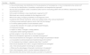
A total of 14 clinical questions were formulated using the PICO format (Patient, Intervention, Comparison, Outcome), grouped by topic, with each question potentially including multiple sub-questions. Table 1 illustrates the clinical questions.
Clinical questions in PICO format.
| Number | Question |
|---|---|
| 1 | a. How should proper risk stratification for the development of hemangioma or risk of complications be carried out? |
| b. How can the identification of possible complications and sequelae be improved? | |
| 2 | Are the HSS and IHReS scales considered useful tools for evaluating patient risk and referral, respectively? When should they be used? |
| 3 | Is the scalp considered a new classification segment for IH? |
| 4 | What tools are currently available for the diagnosis of IH? |
| 5 | What is the utility of imaging modalities in the diagnosis of IH? |
| 6 | In patients with PHACE syndrome, what is the safety profile of the various treatments? |
| 7 | a. Among current treatment options, what is the first-line therapy? |
| b. For which patient profiles is each treatment used? | |
| c. Include the indication for surgery and light-based therapies. | |
| d. Propranolol dosage. | |
| e. Impact on sleep. Changes in sleep patterns. | |
| f. Evaluation before starting treatment. | |
| g. Early surgery or only when there is risk of sequelae? | |
| 8 | What is the therapeutic approach to ulcerated hemangioma? |
| 9 | How should treatment be managed in low-birth-weight and premature infants? |
| 10 | a. Is it possible to extend propranolol treatment beyond 6 months? |
| b. Can treatment be initiated in patients older than 5 months? | |
| 11 | How should sequelae from laser or surgical treatments be managed? |
| 12 | a & b. How should patient monitoring be conducted? |
| c. What are the advantages of telematic monitoring in IH patients? | |
| 13 | How is thermography used to monitor treatment response in IH? |
| 14 | What benefits does family health education provide for IH patients? |
IH: infantile hemangioma; HSS: Hemangioma Severity Scale; IHReS: Infantile Hemangioma Referral Score; PHACE: posterior fossa malformations, hemangioma, arterial anomalies, coarctation of the aorta/cardiac defects, and eye abnormalities.
A stepwise search was conducted to identify relevant national and international publications with clear methodology addressing the selected questions. Publication date restrictions were applied, selecting only studies published after the 2016 consensus document. Only articles published in English or Spanish were included.
First, national and international clinical practice guidelines (CPGs) were identified. Second, the search focused on systematic reviews (SRs) that addressed the defined questions. When evidence was insufficient, primary studies, randomized controlled trials (RCTs), and observational studies were also considered. Databases used included UpToDate (Society guideline links), PubMed (Medline), CENTRAL (Cochrane Central Register of Controlled Trials), and TRIP database.
Study selection was carried out in 2 screening phases: first by title and abstract, then by full text. One reviewer independently assessed the studies for eligibility, with any discrepancies resolved by a second reviewer. References were managed using the Rayyan QCRI online software.
Qualitative research and development of recommendationsAn in-person meeting was held where clinical experts discussed the available evidence, responded to the posed questions, and developed a set of recommendations for managing patients with IH.
Agreement and disagreement percentages for each recommendation were calculated using the Likert scale.9 Consensus was defined as ≥80% agreement.10,11 A 100% agreement was considered unanimity. Agreement between 66% and 79% was considered discrepancy, and <66%, rejection.
The level of evidence and grade of recommendation were evaluated using the 2019 SIGN (Scottish Intercollegiate Guidelines Network) clinical practice guidelines.12
ResultsA total of 107 publications were identified. Of these, 7 were excluded because: (a) they did not answer the defined questions; (b) they involved animal studies; and (c) they were not within the scope of the document. The list of included articles is shown in Table A.1.
Table 2 illustrates the recommendations, including the level of evidence, grade of recommendation, and level of agreement for each.
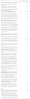
Recommendations from the in-person meeting.
| Recommendation | Level of evidence | Grade of recommendation | Degree of agreement |
|---|---|---|---|
| 1a. For a correct assessment of the increased risk of hemangiomas in a newborn, the following factors should be considered:- Female sex- White race- Prematurity- Low birth weight- Advanced maternal age- Multiple gestation pregnancy- Placenta previa and preeclampsia- Progesterone therapy- Use of assisted reproductive technology or invasive procedures, such as chorionic villus biopsy | 2++ | B | Unanimity |
| 1b. To improve the identification of possible complications and sequelae, the following is recommended:- Early evaluation (before 4 weeks of life) to monitor the hemangioma(s) progression and stratify risk.- Assess each lesion separately.- Conduct reviews at each visit.Supplementary studies may also be necessary to determine associated structural anomalies, such as echocardiogram, hepatic or cerebral ultrasound, cerebral, abdominal, or pelvic magnetic resonance imaging (MRI), ophthalmological examination, and analytical studies to determine thyroid hormones. | 4 | D | Unanimity |
| 2. The HSS and IHReS scales are validated and useful tools for the initial assessment of an IH:- The HSS scale helps determine the initial severity of the hemangioma, including its impact on quality of life and the need to initiate treatment.- The IHReS scale is useful for patient referral by non-expert clinicians. | 2+ | C | Unanimity |
| 3. Considering segmental scalp hemangiomas as a new classification segment for IH, alongside the already known S1 to S4 segments, is recommended. In patients with segmental scalp IH, PHACE syndrome should be ruled out. | 4 | D | Unanimity |
| 4. The currently available tools for IH diagnosis are:- IH diagnosis is clinical, based on medical history and physical examination, recognizing its characteristic appearance.- In cases of uncertain diagnosis, or if an evaluation of the extent is necessary, imaging modalities of the lesion are recommended.- In atypical cases, a biopsy may be performed to rule out other conditions. | 2+ | C | Unanimity |
| 5. Ultrasound is the imaging modality of choice in cases of diagnostic doubt or to determine the extent, which is more frequent in deep, parotid, orbital, or periorbital IHs. Orbital IHs may require MRI to determine their deep extension. Imaging tests are necessary in the following contexts to rule out associated anomalies:- Patients with >5 IHs: abdominal ultrasound.- Patients with segmental hemangioma with a diameter ≥5cm on the face, neck, or scalp: cerebral and cervical MRA, echocardiography, skull MRI with contrast.- Patients with lumbosacral, perineal, or gluteal segmental hemangioma: perform renal ultrasound and lumbosacral spinal MRI to rule out occult spinal dysraphism (in infants younger than 3–4 months, lumbosacral ultrasound may be considered as an alternative).- Midline lumbosacral or sacrococcygeal segmental hemangiomas: spinal MRI. Since it's not an invasive test, pelvic–bladder ultrasound may also be considered.- Segmental hemangiomas of the pelvic region: pelvic/bladder ultrasound. | 2+ | C | Unanimity |
| 6. Beta-blocker treatment is considered safe for children with PHACE syndrome. The lowest possible dose of oral propranolol should be used.The initial dose is 0.5 or 1mg/kg/day within week 1, progressively increasing up to 2 or a maximum of 3mg/kg/day. The optimal dose is reached slowly to minimize abrupt changes in blood pressure. | 2+ | D | Unanimity |
| 7a. For IHs with an absolute indication for treatment, oral propranolol is the first-line therapy. | 1++ | A | Unanimity |
| 7b. Oral corticosteroids (prednisone or prednisolone) would be indicated if beta-blockers cannot be administered.Oral atenolol or nadololwould be alternatives in case of propranolol intolerance.Topical timolol can be prescribed for small and/or superficial IHs. | 1++ | A | Unanimity |
| 7c. Light source therapy may be beneficial for unresponsive ulcerated lesions and for IHs with sequelae.Surgery can be effective for localized ulcerated lesions and lesions with esthetic implications. Surgical excision of the IH is evaluated on an individualized basis. | 2++ | B | Unanimity |
| 7d. The approved oral propranolol dose is 1–3mg/kg per day. The decision must be made based on clinical judgment. | 1++ | A | Unanimity |
| 7e. Parents and family members should be informed that sleep disturbances are the most common adverse effects of propranolol. Adjusting the timing of treatment administration can help mitigate these effects.Severe sleep disturbances can lead to medication discontinuation and affect the quality of life of patients and parents. | 4 | D | Unanimity |
| 7f. Before starting oral propranolol treatment, it's recommended to determine blood pressure, heart rate, and perform cardiopulmonary auscultation. | 2+ | C | Unanimity |
| 7g. The decision for surgery in the proliferative phase depends on exceptional situations (clinical type and location of the IH, and specific symptoms [pain and bleeding]). | 4 | D | Unanimity |
| 8. The treatment of ulcerated IHs includes four main aspects:- Local wound care- Pain reduction- Proliferation reduction- Treatment in case of superinfectionIt's recommended to start treatment for ulcerated IH as soon as possible with local care (non-adhesive dressings and/or barrier creams±topical antibiotic) and pain management.When oral propranolol is indicated, it's preferable to start treatment at doses<1mg/kg/day and slowly escalate the dose (from ≤1mg/kg/day to 2mg/kg/day). Light sources are often used as an adjuvant.For some ulcerated hemangiomas that don’t respond to propranolol or light source therapy, other therapeutic options may be needed, such as adding oral corticosteroids or performing surgical interventions. | 3 | D | Unanimity |
| 9. Premature infants are the group with the highest incidence rate, highest risk of treatment side effects, and also the least studied.There's no established evidence-based treatment for IH in premature newborns.The therapeutic approach in these patients should be cautious and closely monitored.Special precautions should be taken with treatments, and monitoring should be emphasized. Patients should be referred to a specialist for treatment.Oral propranolol is generally used. Propranolol initiation, maintenance, and incremental dosing regimens should be considered. Use with caution in infants younger than 5 weeks corrected age in cases of high significance and start treatment early under close monitoring.In this subgroup, during propranolol treatment, it's necessary to monitor vital signs and start with a dose of 0.5mg/kg/day. It's probably safer to use an initial propranolol dose of 0.5mg/kg/day and admit the infant for at least 2–4h at the start of treatment and with dose increases. Diarrhea and weight loss, in addition to bradycardia and blood pressure, should be carefully controlled.In case of intolerance or undesirable effects to propranolol, atenolol or corticosteroids could be attempted as alternatives.More safety studies are needed for the use of topical beta-blockers in premature infants. | |||
| 10a. Propranolol treatment can be extended beyond 6 months, reaching up to 12 or even, exceptionally, more than 24 months.There's a higher risk of regrowth in patients whose treatment was stopped before 12 months of age (especially before 9 months), and the lowest risk was in those whose treatment was stopped between 12 and 15 months of age. | 2+ | C | Unanimity |
| 10b. Introducing propranolol after the proliferative phase, and even >1 year, appears to be safe and effective.Keep in mind that propranolol treatment is more effective if started during the hemangioma's growth phase. The earlier it's started, the better the functional and/or esthetic results, and it also helps prevent complications. | 2+ | C | Unanimity |
| 11. The management of sequelae from laser or surgical treatments should be approached as follows:- Be aware that some sequelae may improve spontaneously after hemangioma treatment or even with watchful waiting, but invasive procedures, including light sources or surgery, might be needed for satisfactory esthetic results.- Facial hemangiomas are most likely to require surgical treatment or light sources due to esthetic considerations.- Deformity from adipose residue and scarring from ulceration are corrected with surgery and light sources.- Telangiectasia requires treatment with light sources.- Preventing and early recognizing sequelae should be a priority. | 2++ | B | Unanimity |
| 12a. Patient monitoring should include evaluating any adverse events or side effects. It's important to monitor heart rate due to the risk of bradycardia and gastrointestinal symptoms. 12b. During the proliferative stage, clinical follow-up is recommended monthly, then every 3 months.In the involutive phase, reviews can be spaced out more. More frequent follow-up might be needed if there's ulceration or an increase in lesion size. Dynamic infrared (IR) thermography can be used for patient monitoring:- It can track treatment response and detect changes before they’re visible.- Thermal distribution maps can be used for specific local treatments.- It can help identify patients who aren’t responding and monitor individual progress. | 3 | D | Unanimity |
| 12c. Telemedicine/virtual consultation allows for:- An early assessment of pediatric cases.- Reduces the age of treatment initiation in infants with IH.- Rapid referral to outpatient subspecialists for monitoring.- Helps reduce emergency room visitsfor patient monitoring.- Offers a comfortable and remote consultation mode, providing a more accessible and less cumbersome treatment plan, reducing travel and time.- Rapid selection of critical cases in underserved areas, increasing vulnerable patients’ access to high-value care.- Optimizes outcomes by preventing complications and improving treatment initiation. | 3 | D | Consensus |
| 13. Thermography can be used in the outpatient setting for the evaluation and follow-up of IHs. | 3 | D | Unanimity |
| 14. It is recommended to educate families about the nature of IHs, including the expected natural history, potential complications, sequelae, treatment indications, and their adverse effects (sleep disturbances, risk of hypoglycemia, bronchospasm, bradycardia, hypotension, etc.).Formal educational efforts can reduce parental anxiety and improve the management of IH in case of any unexpected situations or worrying changes.The information provided by clinicians should be as specific as possible for the patient's IH.It should be informed if it is low-risk and, therefore, likely not to cause problems or sequelae, or if it is potentially high-risk and requires emergency evaluation or treatment.In infants treated with propranolol, the family should be instructed to recognize the signs of hypoglycemia in infants (hypotonia, hypoactivity, cold sweat). Excessive spacing of feedings should not be allowed.The family should be instructed on the correct administration of the drug and on recognizing signs of hypoglycemia. | 4 | D | Unanimity |
MRA: Magnetic Resonance Angiography; IH: infantile hemangioma; HSS: Hemangioma Severity Scale; IHReS: Infantile Hemangioma Referral Score; IR: infrared; PHACE: posterior fossa malformations, hemangioma, arterial anomalies, coarctation of the aorta/cardiac defects, and eye abnormalities; MRI: magnetic resonance imaging.
The management of IH requires a multidisciplinary approach3 based on early diagnosis, risk stratification, appropriate treatment, and close monitoring.
Diagnosis of IHAs recommended by the scientific committee, the literature indicates that IH diagnosis is generally based on medical history and physical examination.8,13 In cases of diagnostic uncertainty, such as with deep hemangiomas, Doppler ultrasound is usually sufficient.14 Imaging modalities such as magnetic resonance imaging (MRI) may also be necessary to determine depth or extent.15 Additionally, further evaluations are sometimes needed to rule out structural anomalies, such as PHACE or LUMBAR syndromes. These tools not only help confirm the diagnosis but also assess treatment response and IH extent.
Segmental facial IHs were previously categorized into 4 distribution patterns: S1 (frontotemporal segment), S2 (maxillary segment), S3 (mandibular segment), and S4 (frontonasal segment).16 The scientific committee has agreed that scalp IHs constitute a new classification segment. IHs of the face, scalp, and neck are associated with PHACE syndrome6; thus, experts recommend ruling out PHACE in patients with segmental scalp IHs.
Risk stratification of IHRisk stratification is essential to prevent severe complications and determine the most appropriate treatment.7 The previous consensus defined high-risk hemangiomas as those located on the face or lumbosacral area with a diameter>5cm, or ulcerated IHs.5 For this update, the committee indicated that 2 scales can be used to evaluate severity: the Hemangioma Severity Scale (HSS)17 and the Infantile Hemangioma Referral Score (IHReS).18 The former allows precise risk evaluation, while the latter is a validated referral tool developed by expert panels and tested by pediatricians and general practitioners to improve referral decision-making.18 The 2 scales enable timely and appropriate intervention based on the patient's risk profile.
The 2016 consensus document emphasized the psychological impact of potential sequelae.5 In this update, early assessment and regular follow-up are recommended, as well as the use of additional imaging modalities or lab tests to identify and prevent sequelae. Addressing sequelae of facial IHs is especially important and may require laser or surgical correction of deformities or scars.
Treatment of IHOral propranolol remains the first-line therapy for IH,4,19 with a recommended dose of 1–3mg/kg/day.19,20 The expert panel recommends that dose adjustment be made based on clinical judgment. Before initiating propranolol, assessment of blood pressure, heart rate, and cardiopulmonary auscultation is advised.
Beta-blocker treatment with propranolol has demonstrated to be safe and effective, even in patients with PHACE syndrome.6,19,21,22 The committee recommends starting at the lowest possible dose (0.5 or 1mg/kg/day), gradually up titrating to 2 or 3mg/kg/day for PHACE patients.
Although the incidence rate of IH is more common in low-birth-weight and preterm infants,23,24 no evidence-based treatment protocol has been established for this population.24 However, some data indicate that propranolol is also safe and effective in low-weight and premature patients.25,26 The scientific committee emphasizes the importance of close monitoring and follow-up for this group. Starting, maintenance, and incremental propranolol dosing regimens should be carefully planned. Oral propranolol should be used cautiously and only when clearly needed, especially in infants with corrected age<5 weeks, with treatment initiated early under close monitoring.
Both data and the scientific committee suggest that although the standard course of propranolol treatment is 6 months, it may be extended to 12 or even 24 months—or longer in specific cases depending on lesion type and response.27–31 Some studies indicate that prolonged treatment is associated with a lower relapse rate.13
A very common adverse effect of propranolol is sleep disturbance.32 Experts recommend informing families and adjusting the timing of administration to mitigate these effects. Administering the first dose early in the morning and the second dose 8h later may reduce sleep disruption. Oral propranolol should always be administered after feedings to avoid hypoglycemia.
Other treatment options include oral corticosteroids in patients for whom beta-blockers are contraindicated—e.g. (a) potential drug interactions; (b) sick sinus syndrome; (c) bradycardia or hypotension; (d) heart failure; (e) predisposition to hypoglycemia; (f) hypersensitivity to propranolol hydrochloride; (g) pheochromocytoma; or (h) Raynaud's syndrome.13 Topical timolol can be used for thin, superficial IHs,8 and atenolol is an alternative to propranolol.33 Surgery and light-based therapy are also recommended for residual lesions.34 Surgery may be considered in specific cases such as ulcerated or bleeding IHs, or where complications or sequelae are likely.35
Monitoring of patients with IHMonitoring is essential to assess treatment response36 and detect adverse effects.30 Follow-up should be periodic and include evaluation of vital signs (e.g., heart rate),30 GI symptoms,19 and sleep disturbances.37 Experts highlight the advantages of infrared thermography (IR) for IH monitoring, as it is a non-invasive, contact-free, and cost-effective technique.38
The scientific committee recommends an initial clinical follow-up at 1 month and then every 3 months until involution. More frequent follow-up may be necessary in cases of ulceration or lesion growth.
Telemedicine is also highlighted as a monitoring option to reduce travel needs and improve care access.
The scientific committee values the importance of family health education in IH management, as it helps reduce family anxiety and supports more efficient care.
Study limitationsThis update of the 2016 IH consensus is based on existing literature and the clinical experience of an expert panel from across Spain. The results may not be generalizable outside of Spain. Moreover, implementation of the recommendations may vary depending on the resources available in each center.
ConclusionsThis update of the IH consensus document introduces several changes vs the 2016 version: (a) segmental scalp hemangiomas are now defined as a new IH classification segment; (b) early referral of IH patients to specialists using the IHReS tool is emphasized; (c) the safety of beta-blocker treatment (e.g., propranolol) in PHACE syndrome is reaffirmed; (d) first-line propranolol therapy may be extended beyond 6 months, up to 12 or 24 months in exceptional cases, since early discontinuation is associated with higher risk of regrowth; and (e) the advantages of implementing telemedicine for IH follow-up are emphasized, including convenience, earlier evaluation, better optimization of treatment initiation, improved outcomes, and prevention of IH-related complications.
FundingThis study was funded by Laboratorios Pierre Fabre S.A.
Conflicts of interestEB declares financial and administrative support, article publication fees, travel, and writing assistance from Pierre Fabre Dermatology; also to keep a relationship with ISSVA including board membership, Sanofi, LEO Pharma, Almirall S.A., Viatris, ISDIN, and Novartis Pharmaceuticals Corporation including consultancy, speaking fees, and/or travel reimbursement. JBW declares no financial interests or any personal relationships that could have influenced the work reported in this article. IBM declares no financial interests or personal relationships that could have influenced the work reported. MCD reports financial support from Pierre Fabre S.A., and a relationship with the company including funding, speaker fees, and travel reimbursement. ACS reports financial, administrative, publication, and writing support from Pierre Fabre S.A. JB reports financial and writing support from Pierre Fabre S.A. RLL reports financial, administrative, and writing support from Pierre Fabre S.A. JPL declares no financial interests or personal relationships that could have influenced the work. AHM reports financial, administrative, publication, statistical analysis, and writing support from Laboratoires Pierre Fabre; and relationships with Sanofi S.A. and Viatris (consulting, speaker fees), LETI Pharma GmbH and Beiersdorf SA (speaker fees), and the European Society for Pediatric Dermatology (ESPD, board membership, travel reimbursement). LJM declares no financial interests or personal relationships that could have influenced the work. JCLG reports financial support from Pierre Fabre S.A. and a relationship including speaking fees. AMS reports financial support from Pierre Fabre S.A., including funding, speaker fees, and travel reimbursement. PR reports financial support from Pierre Fabre. ATF reports writing assistance from Laboratoires Pierre Fabre. AVV reports having received consultancy fees and/or honoraria for speaking, presenting, manuscript writing, or attending meetings from Amryt, Lilly, AbbVie, Almirall, Amgen, Boehringer Ingelheim, Bristol-Myers Squibb, Ferrer, Galderma, Novartis, Pierre Fabre, Pfizer, Sanofi Genzyme, and Jansen.
The authors thank Laboratorios Pierre Fabre S.A. for funding this research, and GOC for methodological and manuscript writing support.
The followings are the supplementary data to this article:


