array:23 [
  "pii" => "S0001731023001618"
  "issn" => "00017310"
  "doi" => "10.1016/j.ad.2023.02.009"
  "estado" => "S300"
  "fechaPublicacion" => "2023-04-01"
  "aid" => "3407"
  "copyrightAnyo" => "2023"
  "documento" => "simple-article"
  "crossmark" => 1
  "subdocumento" => "crp"
  "cita" => "Actas Dermosifiliogr. 2023;114:T343-T344"
  "abierto" => array:3 [
    "ES" => true
    "ES2" => true
    "LATM" => true
  ]
  "gratuito" => true
  "lecturas" => array:1 [
    "total" => 0
  ]
  "itemSiguiente" => array:18 [
    "pii" => "S0001731022010602"
    "issn" => "00017310"
    "doi" => "10.1016/j.ad.2022.03.032"
    "estado" => "S300"
    "fechaPublicacion" => "2023-04-01"
    "aid" => "3344"
    "copyright" => "AEDV"
    "documento" => "simple-article"
    "crossmark" => 1
    "subdocumento" => "crp"
    "cita" => "Actas Dermosifiliogr. 2023;114:345-6"
    "abierto" => array:3 [
      "ES" => true
      "ES2" => true
      "LATM" => true
    ]
    "gratuito" => true
    "lecturas" => array:1 [
      "total" => 0
    ]
    "es" => array:11 [
      "idiomaDefecto" => true
      "cabecera" => "<span class="elsevierStyleTextfn">CASOS PARA EL DIAGN&#211;STICO</span>"
      "titulo" => "Manchas de color marr&#243;n-rojizo en las palmas de un ni&#241;o"
      "tienePdf" => "es"
      "tieneTextoCompleto" => "es"
      "paginas" => array:1 [
        0 => array:2 [
          "paginaInicial" => "345"
          "paginaFinal" => "346"
        ]
      ]
      "titulosAlternativos" => array:1 [
        "en" => array:1 [
          "titulo" => "Red-Brown Patches on a Boy&#39;s Palms"
        ]
      ]
      "contieneTextoCompleto" => array:1 [
        "es" => true
      ]
      "contienePdf" => array:1 [
        "es" => true
      ]
      "resumenGrafico" => array:2 [
        "original" => 0
        "multimedia" => array:6 [
          "identificador" => "fig0005"
          "etiqueta" => "Figura 1"
          "tipo" => "MULTIMEDIAFIGURA"
          "mostrarFloat" => true
          "mostrarDisplay" => false
          "figura" => array:1 [
            0 => array:4 [
              "imagen" => "gr1.jpeg"
              "Alto" => 905
              "Ancho" => 755
              "Tamanyo" => 127302
            ]
          ]
        ]
      ]
      "autores" => array:1 [
        0 => array:2 [
          "autoresLista" => "A&#46;A&#46; Gonz&#225;lez Ruiz, A&#46; Bot&#237;a Paco, A&#46; Docampo Sim&#243;n, M&#46; Niveiro de Jaime, I&#46; Betlloch-Mas"
          "autores" => array:5 [
            0 => array:2 [
              "nombre" => "A&#46;A&#46;"
              "apellidos" => "Gonz&#225;lez Ruiz"
            ]
            1 => array:2 [
              "nombre" => "A&#46;"
              "apellidos" => "Bot&#237;a Paco"
            ]
            2 => array:2 [
              "nombre" => "A&#46;"
              "apellidos" => "Docampo Sim&#243;n"
            ]
            3 => array:2 [
              "nombre" => "M&#46;"
              "apellidos" => "Niveiro de Jaime"
            ]
            4 => array:2 [
              "nombre" => "I&#46;"
              "apellidos" => "Betlloch-Mas"
            ]
          ]
        ]
      ]
    ]
    "idiomaDefecto" => "es"
    "EPUB" => "https://multimedia.elsevier.es/PublicationsMultimediaV1/item/epub/S0001731022010602?idApp=UINPBA000044"
    "url" => "/00017310/0000011400000004/v1_202304111712/S0001731022010602/v1_202304111712/es/main.assets"
  ]
  "itemAnterior" => array:17 [
    "pii" => "S0001731022006767"
    "issn" => "00017310"
    "doi" => "10.1016/j.ad.2021.07.028"
    "estado" => "S300"
    "fechaPublicacion" => "2023-04-01"
    "aid" => "3159"
    "documento" => "simple-article"
    "crossmark" => 1
    "subdocumento" => "crp"
    "cita" => "Actas Dermosifiliogr. 2023;114:343-4"
    "abierto" => array:3 [
      "ES" => true
      "ES2" => true
      "LATM" => true
    ]
    "gratuito" => true
    "lecturas" => array:1 [
      "total" => 0
    ]
    "es" => array:11 [
      "idiomaDefecto" => true
      "cabecera" => "<span class="elsevierStyleTextfn">CASOS PARA EL DIAGN&#211;STICO</span>"
      "titulo" => "P&#225;pula pedunculada en regi&#243;n plantar"
      "tienePdf" => "es"
      "tieneTextoCompleto" => "es"
      "paginas" => array:1 [
        0 => array:2 [
          "paginaInicial" => "343"
          "paginaFinal" => "344"
        ]
      ]
      "titulosAlternativos" => array:1 [
        "en" => array:1 [
          "titulo" => "Pedunculated Papule on the Sole of the Foot"
        ]
      ]
      "contieneTextoCompleto" => array:1 [
        "es" => true
      ]
      "contienePdf" => array:1 [
        "es" => true
      ]
      "resumenGrafico" => array:2 [
        "original" => 0
        "multimedia" => array:6 [
          "identificador" => "fig0005"
          "etiqueta" => "Figura 1"
          "tipo" => "MULTIMEDIAFIGURA"
          "mostrarFloat" => true
          "mostrarDisplay" => false
          "figura" => array:1 [
            0 => array:4 [
              "imagen" => "gr1.jpeg"
              "Alto" => 1586
              "Ancho" => 893
              "Tamanyo" => 119476
            ]
          ]
        ]
      ]
      "autores" => array:1 [
        0 => array:2 [
          "autoresLista" => "I&#46; Arregui, C&#46; &#209;anco, F&#46; Mardones"
          "autores" => array:3 [
            0 => array:2 [
              "nombre" => "I&#46;"
              "apellidos" => "Arregui"
            ]
            1 => array:2 [
              "nombre" => "C&#46;"
              "apellidos" => "&#209;anco"
            ]
            2 => array:2 [
              "nombre" => "F&#46;"
              "apellidos" => "Mardones"
            ]
          ]
        ]
      ]
    ]
    "idiomaDefecto" => "es"
    "EPUB" => "https://multimedia.elsevier.es/PublicationsMultimediaV1/item/epub/S0001731022006767?idApp=UINPBA000044"
    "url" => "/00017310/0000011400000004/v1_202304111712/S0001731022006767/v1_202304111712/es/main.assets"
  ]
  "asociados" => array:1 [
    0 => array:17 [
      "pii" => "S0001731022006767"
      "issn" => "00017310"
      "doi" => "10.1016/j.ad.2021.07.028"
      "estado" => "S300"
      "fechaPublicacion" => "2023-04-01"
      "aid" => "3159"
      "documento" => "simple-article"
      "crossmark" => 1
      "subdocumento" => "crp"
      "cita" => "Actas Dermosifiliogr. 2023;114:343-4"
      "abierto" => array:3 [
        "ES" => true
        "ES2" => true
        "LATM" => true
      ]
      "gratuito" => true
      "lecturas" => array:1 [
        "total" => 0
      ]
      "es" => array:11 [
        "idiomaDefecto" => true
        "cabecera" => "<span class="elsevierStyleTextfn">CASOS PARA EL DIAGN&#211;STICO</span>"
        "titulo" => "P&#225;pula pedunculada en regi&#243;n plantar"
        "tienePdf" => "es"
        "tieneTextoCompleto" => "es"
        "paginas" => array:1 [
          0 => array:2 [
            "paginaInicial" => "343"
            "paginaFinal" => "344"
          ]
        ]
        "titulosAlternativos" => array:1 [
          "en" => array:1 [
            "titulo" => "Pedunculated Papule on the Sole of the Foot"
          ]
        ]
        "contieneTextoCompleto" => array:1 [
          "es" => true
        ]
        "contienePdf" => array:1 [
          "es" => true
        ]
        "resumenGrafico" => array:2 [
          "original" => 0
          "multimedia" => array:6 [
            "identificador" => "fig0005"
            "etiqueta" => "Figura 1"
            "tipo" => "MULTIMEDIAFIGURA"
            "mostrarFloat" => true
            "mostrarDisplay" => false
            "figura" => array:1 [
              0 => array:4 [
                "imagen" => "gr1.jpeg"
                "Alto" => 1586
                "Ancho" => 893
                "Tamanyo" => 119476
              ]
            ]
          ]
        ]
        "autores" => array:1 [
          0 => array:2 [
            "autoresLista" => "I&#46; Arregui, C&#46; &#209;anco, F&#46; Mardones"
            "autores" => array:3 [
              0 => array:2 [
                "nombre" => "I&#46;"
                "apellidos" => "Arregui"
              ]
              1 => array:2 [
                "nombre" => "C&#46;"
                "apellidos" => "&#209;anco"
              ]
              2 => array:2 [
                "nombre" => "F&#46;"
                "apellidos" => "Mardones"
              ]
            ]
          ]
        ]
      ]
      "idiomaDefecto" => "es"
      "EPUB" => "https://multimedia.elsevier.es/PublicationsMultimediaV1/item/epub/S0001731022006767?idApp=UINPBA000044"
      "url" => "/00017310/0000011400000004/v1_202304111712/S0001731022006767/v1_202304111712/es/main.assets"
    ]
  ]
  "en" => array:14 [
    "idiomaDefecto" => true
    "cabecera" => "<span class="elsevierStyleTextfn">Cases for Diagnosis</span>"
    "titulo" => " Pedunculated Papule on the Sole of the Foot"
    "tieneTextoCompleto" => true
    "paginas" => array:1 [
      0 => array:2 [
        "paginaInicial" => "T343"
        "paginaFinal" => "T344"
      ]
    ]
    "autores" => array:1 [
      0 => array:4 [
        "autoresLista" => "I&#46; Arregui, C&#46; &#209;anco, F&#46; Mardones"
        "autores" => array:3 [
          0 => array:4 [
            "nombre" => "I&#46;"
            "apellidos" => "Arregui"
            "email" => array:1 [
              0 => "isidora.arregui@gmail.com"
            ]
            "referencia" => array:2 [
              0 => array:2 [
                "etiqueta" => "<span class="elsevierStyleSup">a</span>"
                "identificador" => "aff0005"
              ]
              1 => array:2 [
                "etiqueta" => "<span class="elsevierStyleSup">&#42;</span>"
                "identificador" => "cor0005"
              ]
            ]
          ]
          1 => array:3 [
            "nombre" => "C&#46;"
            "apellidos" => "&#209;anco"
            "referencia" => array:1 [
              0 => array:2 [
                "etiqueta" => "<span class="elsevierStyleSup">b</span>"
                "identificador" => "aff0010"
              ]
            ]
          ]
          2 => array:3 [
            "nombre" => "F&#46;"
            "apellidos" => "Mardones"
            "referencia" => array:1 [
              0 => array:2 [
                "etiqueta" => "<span class="elsevierStyleSup">c</span>"
                "identificador" => "aff0015"
              ]
            ]
          ]
        ]
        "afiliaciones" => array:3 [
          0 => array:3 [
            "entidad" => "M&#233;dico cirujano&#44; Universidad de los Andes&#44; Santiago&#44; Chile"
            "etiqueta" => "a"
            "identificador" => "aff0005"
          ]
          1 => array:3 [
            "entidad" => "Residente Dermatolog&#237;a 2&#176; a&#241;o&#44; Universidad de los Andes&#44; Santiago&#44; Chile"
            "etiqueta" => "b"
            "identificador" => "aff0010"
          ]
          2 => array:3 [
            "entidad" => "Dermat&#243;logo&#44; Cl&#237;nica Universidad de los Andes&#44; Santiago&#44; Chile"
            "etiqueta" => "c"
            "identificador" => "aff0015"
          ]
        ]
        "correspondencia" => array:1 [
          0 => array:3 [
            "identificador" => "cor0005"
            "etiqueta" => "&#8270;"
            "correspondencia" => "Corresponding author&#46;"
          ]
        ]
      ]
    ]
    "titulosAlternativos" => array:1 [
      "es" => array:1 [
        "titulo" => "P&#225;pula pedunculada en regi&#243;n plantar"
      ]
    ]
    "resumenGrafico" => array:2 [
      "original" => 0
      "multimedia" => array:7 [
        "identificador" => "fig0010"
        "etiqueta" => "Figure 2"
        "tipo" => "MULTIMEDIAFIGURA"
        "mostrarFloat" => true
        "mostrarDisplay" => false
        "figura" => array:1 [
          0 => array:4 [
            "imagen" => "gr2.jpeg"
            "Alto" => 569
            "Ancho" => 756
            "Tamanyo" => 191083
          ]
        ]
        "descripcion" => array:1 [
          "en" => "<p id="spar0005" class="elsevierStyleSimplePara elsevierViewall">Hematoxylin&#8211;eosin&#44; 200&#215;&#46;</p>"
        ]
      ]
    ]
    "textoCompleto" => "<span class="elsevierStyleSections"><span id="sec0005" class="elsevierStyleSection elsevierViewall"><span class="elsevierStyleSectionTitle" id="sect0005">Medical History</span><p id="par0005" class="elsevierStylePara elsevierViewall">An otherwise healthy 45-year-old man consulted for an asymptomatic lesion on the middle third of the right sole that had first appeared 18 months earlier&#46; The patient did not recall injury to the area or the presence of a previous lesion&#46;</p></span><span id="sec0010" class="elsevierStyleSection elsevierViewall"><span class="elsevierStyleSectionTitle" id="sect0010">Physical Examination</span><p id="par0010" class="elsevierStylePara elsevierViewall">Physical examination revealed a flesh-colored pedunculated tumor measuring 8<span class="elsevierStyleHsp" style=""></span>mm in diameter&#46; The tumor was soft&#44; with a smooth surface&#44; and was located on the middle third of the right sole &#40;<a class="elsevierStyleCrossRef" href="#fig0005">Fig&#46; 1</a>&#41;&#46;</p><elsevierMultimedia ident="fig0005"></elsevierMultimedia></span><span id="sec0015" class="elsevierStyleSection elsevierViewall"><span class="elsevierStyleSectionTitle" id="sect0015">Histopathology</span><p id="par0015" class="elsevierStylePara elsevierViewall">The tumor was removed using excisional biopsy&#46; Histopathology revealed a proliferation of spindle-shaped homogeneous cells immersed in a myxoid stroma in the dermis &#40;<a class="elsevierStyleCrossRef" href="#fig0010">Fig&#46; 2</a>&#44; hematoxylin&#8211;eosin&#44; 200&#215;&#41;&#46; The results of immunohistochemistry were positive for CD34 and negative for S100 &#40;<a class="elsevierStyleCrossRef" href="#fig0015">Fig&#46; 3</a>&#44; immunohistochemistry CD34&#44; 200&#215;&#41;&#46;</p><elsevierMultimedia ident="fig0010"></elsevierMultimedia><elsevierMultimedia ident="fig0015"></elsevierMultimedia><p id="par0020" class="elsevierStylePara elsevierViewall">What is your diagnosis&#63;</p></span><span id="sec0020" class="elsevierStyleSection elsevierViewall"><span class="elsevierStyleSectionTitle" id="sect0020">Diagnosis</span><p id="par0025" class="elsevierStylePara elsevierViewall">Superficial acral fibromyxoma&#46;</p></span><span id="sec0025" class="elsevierStyleSection elsevierViewall"><span class="elsevierStyleSectionTitle" id="sect0025">Clinical Course and Treatment</span><p id="par0030" class="elsevierStylePara elsevierViewall">After diagnosis&#44; the surgical margins were extended by 0&#46;5<span class="elsevierStyleHsp" style=""></span>mm&#46; Histology revealed no relevant findings&#46; No signs of recurrence have been observed after 10 months of follow-up&#46;</p></span><span id="sec0030" class="elsevierStyleSection elsevierViewall"><span class="elsevierStyleSectionTitle" id="sect0030">Commentary</span><p id="par0035" class="elsevierStylePara elsevierViewall">Superficial acral fibromyxoma is a benign mesenchymal tumor that mainly affects men in the fifth decade of life&#46; In most published cases&#44; the lesion is found in the periungual and subungual regions&#46;<a class="elsevierStyleCrossRefs" href="#bib0035"><span class="elsevierStyleSup">1&#8211;3</span></a></p><p id="par0040" class="elsevierStylePara elsevierViewall">Clinically&#44; the tumor is usually nonencapsulated&#44; well-circumscribed&#44; polypoid or verrucous&#44; and dome-shaped&#46; It extends throughout the dermis&#44; potentially as far as the subcutaneous tissue&#44; underlying fascia&#44; or periosteal layer&#46;<a class="elsevierStyleCrossRefs" href="#bib0040"><span class="elsevierStyleSup">2&#8211;4</span></a></p><p id="par0045" class="elsevierStylePara elsevierViewall">Radiology findings are nonspecific&#44; although bone erosions or lytic lesions may co-occur in one-third of cases&#46;<a class="elsevierStyleCrossRefs" href="#bib0035"><span class="elsevierStyleSup">1&#8211;5</span></a></p><p id="par0050" class="elsevierStylePara elsevierViewall">Histopathology may reveal hyperkeratosis in the epidermis&#46; Proliferation of spindle-shaped fibroblasts within a myxoid matrix&#44; with nuclear atypia and mast cell infiltration&#44; is a key finding in the dermis&#46; Immunohistochemistry is positive for CD34&#44; CD99&#44; vimentin&#44; and epithelial membrane antigen and negative for neural and muscular differentiation markers &#40;S100&#44; HMB-45&#44; SMA&#44; desmin&#44; and actin&#41;&#46;<a class="elsevierStyleCrossRefs" href="#bib0035"><span class="elsevierStyleSup">1&#44;3&#44;5</span></a></p><p id="par0055" class="elsevierStylePara elsevierViewall">The differential diagnosis includes fibrokeratoma&#44; fibroma&#44; and plantar warts&#46;<a class="elsevierStyleCrossRef" href="#bib0045"><span class="elsevierStyleSup">3</span></a> It is also important to bear in mind tumors that are negative for CD34 &#40;glomus tumor&#44; sclerosing perineurioma&#44; acquired digital fibrokeratoma&#44; and periungual fibroma&#41;&#44; as well as those that are positive for CD34 &#40;superficial angiomyxoma&#44; dermatofibrosarcoma protuberans&#44; and myxoid neurofibroma&#41;&#46;<a class="elsevierStyleCrossRefs" href="#bib0035"><span class="elsevierStyleSup">1&#44;3&#44;5</span></a></p><p id="par0060" class="elsevierStylePara elsevierViewall">The treatment of choice is surgical resection with free margins&#44; since local recurrence is recorded in approximately 20&#37;&#8211;25&#37; of cases&#46; While the natural course of the tumor is benign&#44; the histopathologic spectrum of spindle-shaped fibroblasts is variable&#44; ranging from mild nuclear atypia to major atypia&#44; with transformation to a low-grade malignant tumor being theoretically possible&#46;<a class="elsevierStyleCrossRefs" href="#bib0050"><span class="elsevierStyleSup">4&#44;6</span></a> However&#44; prognosis is excellent&#44; and&#44; to date&#44; there have been no published reports of malignant transformation or metastases&#46;<a class="elsevierStyleCrossRefs" href="#bib0035"><span class="elsevierStyleSup">1&#44;3&#8211;5</span></a></p></span><span id="sec0035" class="elsevierStyleSection elsevierViewall"><span class="elsevierStyleSectionTitle" id="sect0035">Conflicts of Interest</span><p id="par0065" class="elsevierStylePara elsevierViewall">The authors declare that they have no financial or personal relationship with other persons or organizations that could give rise to a conflict of interest with respect to the present article&#46;</p></span></span>"
    "textoCompletoSecciones" => array:1 [
      "secciones" => array:8 [
        0 => array:2 [
          "identificador" => "sec0005"
          "titulo" => "Medical History"
        ]
        1 => array:2 [
          "identificador" => "sec0010"
          "titulo" => "Physical Examination"
        ]
        2 => array:2 [
          "identificador" => "sec0015"
          "titulo" => "Histopathology"
        ]
        3 => array:2 [
          "identificador" => "sec0020"
          "titulo" => "Diagnosis"
        ]
        4 => array:2 [
          "identificador" => "sec0025"
          "titulo" => "Clinical Course and Treatment"
        ]
        5 => array:2 [
          "identificador" => "sec0030"
          "titulo" => "Commentary"
        ]
        6 => array:2 [
          "identificador" => "sec0035"
          "titulo" => "Conflicts of Interest"
        ]
        7 => array:1 [
          "titulo" => "References"
        ]
      ]
    ]
    "pdfFichero" => "main.pdf"
    "tienePdf" => true
    "multimedia" => array:3 [
      0 => array:6 [
        "identificador" => "fig0005"
        "etiqueta" => "Figure 1"
        "tipo" => "MULTIMEDIAFIGURA"
        "mostrarFloat" => true
        "mostrarDisplay" => false
        "figura" => array:1 [
          0 => array:4 [
            "imagen" => "gr1.jpeg"
            "Alto" => 1058
            "Ancho" => 597
            "Tamanyo" => 60961
          ]
        ]
      ]
      1 => array:7 [
        "identificador" => "fig0010"
        "etiqueta" => "Figure 2"
        "tipo" => "MULTIMEDIAFIGURA"
        "mostrarFloat" => true
        "mostrarDisplay" => false
        "figura" => array:1 [
          0 => array:4 [
            "imagen" => "gr2.jpeg"
            "Alto" => 569
            "Ancho" => 756
            "Tamanyo" => 191083
          ]
        ]
        "descripcion" => array:1 [
          "en" => "<p id="spar0005" class="elsevierStyleSimplePara elsevierViewall">Hematoxylin&#8211;eosin&#44; 200&#215;&#46;</p>"
        ]
      ]
      2 => array:7 [
        "identificador" => "fig0015"
        "etiqueta" => "Figure 3"
        "tipo" => "MULTIMEDIAFIGURA"
        "mostrarFloat" => true
        "mostrarDisplay" => false
        "figura" => array:1 [
          0 => array:4 [
            "imagen" => "gr3.jpeg"
            "Alto" => 574
            "Ancho" => 761
            "Tamanyo" => 160612
          ]
        ]
        "descripcion" => array:1 [
          "en" => "<p id="spar0010" class="elsevierStyleSimplePara elsevierViewall">Immunohistochemical staining for CD34&#44; 200&#215;&#46;</p>"
        ]
      ]
    ]
    "bibliografia" => array:2 [
      "titulo" => "References"
      "seccion" => array:1 [
        0 => array:2 [
          "identificador" => "bibs0015"
          "bibliografiaReferencia" => array:6 [
            0 => array:3 [
              "identificador" => "bib0035"
              "etiqueta" => "1"
              "referencia" => array:1 [
                0 => array:2 [
                  "contribucion" => array:1 [
                    0 => array:2 [
                      "titulo" => "Superficial acral fibromyxoma&#44; appearing as retronychia&#58; a rare clinical entity"
                      "autores" => array:1 [
                        0 => array:2 [
                          "etal" => false
                          "autores" => array:6 [
                            0 => "E&#46; Kalloniati"
                            1 => "S&#46; Arampatzi"
                            2 => "C&#46; Ouzouni"
                            3 => "D&#46; Papathemeli"
                            4 => "E&#46; Lazaridou"
                            5 => "M&#46;-G&#46; Trakatelli"
                          ]
                        ]
                      ]
                    ]
                  ]
                  "host" => array:1 [
                    0 => array:2 [
                      "doi" => "10.1159/000507900"
                      "Revista" => array:6 [
                        "tituloSerie" => "Skin Appendage Disord"
                        "fecha" => "2020"
                        "volumen" => "6"
                        "paginaInicial" => "312"
                        "paginaFinal" => "314"
                        "link" => array:1 [
                          0 => array:2 [
                            "url" => "https://www.ncbi.nlm.nih.gov/pubmed/33088818"
                            "web" => "Medline"
                          ]
                        ]
                      ]
                    ]
                  ]
                ]
              ]
            ]
            1 => array:3 [
              "identificador" => "bib0040"
              "etiqueta" => "2"
              "referencia" => array:1 [
                0 => array:2 [
                  "contribucion" => array:1 [
                    0 => array:2 [
                      "titulo" => "Superficial acral fibromyxoma"
                      "autores" => array:1 [
                        0 => array:2 [
                          "etal" => false
                          "autores" => array:2 [
                            0 => "J&#46;L&#46; Sawaya"
                            1 => "A&#46; Khachemoune"
                          ]
                        ]
                      ]
                    ]
                  ]
                  "host" => array:1 [
                    0 => array:2 [
                      "doi" => "10.1111/ijd.12750"
                      "Revista" => array:6 [
                        "tituloSerie" => "Int J Dermatol"
                        "fecha" => "2015"
                        "volumen" => "54"
                        "paginaInicial" => "499"
                        "paginaFinal" => "508"
                        "link" => array:1 [
                          0 => array:2 [
                            "url" => "https://www.ncbi.nlm.nih.gov/pubmed/25772615"
                            "web" => "Medline"
                          ]
                        ]
                      ]
                    ]
                  ]
                ]
              ]
            ]
            2 => array:3 [
              "identificador" => "bib0045"
              "etiqueta" => "3"
              "referencia" => array:1 [
                0 => array:2 [
                  "contribucion" => array:1 [
                    0 => array:2 [
                      "titulo" => "Rare plantar heel presentation of superficial acral fibromyxoma"
                      "autores" => array:1 [
                        0 => array:2 [
                          "etal" => false
                          "autores" => array:5 [
                            0 => "V&#46; Campbell"
                            1 => "N&#46; Machnikowski"
                            2 => "J&#46; Houghton"
                            3 => "B&#46; Murphy"
                            4 => "O&#46; Kerr"
                          ]
                        ]
                      ]
                    ]
                  ]
                  "host" => array:1 [
                    0 => array:2 [
                      "doi" => "10.1111/dth.14517"
                      "Revista" => array:3 [
                        "tituloSerie" => "Dermatol Ther"
                        "fecha" => "2020"
                        "volumen" => "33"
                      ]
                    ]
                  ]
                ]
              ]
            ]
            3 => array:3 [
              "identificador" => "bib0050"
              "etiqueta" => "4"
              "referencia" => array:1 [
                0 => array:2 [
                  "contribucion" => array:1 [
                    0 => array:2 [
                      "titulo" => "Superficial acral fibromyxoma in unusual location&#58; a case report"
                      "autores" => array:1 [
                        0 => array:2 [
                          "etal" => false
                          "autores" => array:5 [
                            0 => "B&#46;E&#46; Crepaldi"
                            1 => "R&#46;D&#46; Soares"
                            2 => "F&#46;D&#46; Silveira"
                            3 => "R&#46;I&#46; Taira"
                            4 => "M&#46;H&#46; Matsumoto"
                          ]
                        ]
                      ]
                    ]
                  ]
                  "host" => array:1 [
                    0 => array:2 [
                      "doi" => "10.1111/ans.15342"
                      "Revista" => array:6 [
                        "tituloSerie" => "ANZ J Surg"
                        "fecha" => "2020"
                        "volumen" => "90"
                        "paginaInicial" => "888"
                        "paginaFinal" => "889"
                        "link" => array:1 [
                          0 => array:2 [
                            "url" => "https://www.ncbi.nlm.nih.gov/pubmed/31334594"
                            "web" => "Medline"
                          ]
                        ]
                      ]
                    ]
                  ]
                ]
              ]
            ]
            4 => array:3 [
              "identificador" => "bib0055"
              "etiqueta" => "5"
              "referencia" => array:1 [
                0 => array:2 [
                  "contribucion" => array:1 [
                    0 => array:2 [
                      "titulo" => "Case for diagnosis&#46; Single-digit clubbing"
                      "autores" => array:1 [
                        0 => array:2 [
                          "etal" => false
                          "autores" => array:4 [
                            0 => "L&#46; Crestani"
                            1 => "I&#46;A&#46; Fasciani"
                            2 => "P&#46; Kakizaki"
                            3 => "N&#46;Y&#46;S&#46; Valente"
                          ]
                        ]
                      ]
                    ]
                  ]
                  "host" => array:1 [
                    0 => array:2 [
                      "doi" => "10.1016/j.abd.2020.01.006"
                      "Revista" => array:6 [
                        "tituloSerie" => "An Bras Dermatol"
                        "fecha" => "2020"
                        "volumen" => "95"
                        "paginaInicial" => "524"
                        "paginaFinal" => "526"
                        "link" => array:1 [
                          0 => array:2 [
                            "url" => "https://www.ncbi.nlm.nih.gov/pubmed/32448714"
                            "web" => "Medline"
                          ]
                        ]
                      ]
                    ]
                  ]
                ]
              ]
            ]
            5 => array:3 [
              "identificador" => "bib0060"
              "etiqueta" => "6"
              "referencia" => array:1 [
                0 => array:2 [
                  "contribucion" => array:1 [
                    0 => array:2 [
                      "titulo" => "Superficial acral fibromyxoma&#58; a clinicopathologic and immunohistochemical analysis of 37 cases of a distinctive soft tissue tumor with a predilection for the fingers and toes"
                      "autores" => array:1 [
                        0 => array:2 [
                          "etal" => false
                          "autores" => array:3 [
                            0 => "J&#46;F&#46; Fetsch"
                            1 => "W&#46;B&#46; Laskin"
                            2 => "M&#46; Miettinen"
                          ]
                        ]
                      ]
                    ]
                  ]
                  "host" => array:1 [
                    0 => array:2 [
                      "doi" => "10.1053/hupa.2001.25903"
                      "Revista" => array:5 [
                        "tituloSerie" => "Human Pathol"
                        "fecha" => "2001"
                        "volumen" => "32"
                        "paginaInicial" => "704"
                        "paginaFinal" => "714"
                      ]
                    ]
                  ]
                ]
              ]
            ]
          ]
        ]
      ]
    ]
  ]
  "idiomaDefecto" => "en"
  "url" => "/00017310/0000011400000004/v1_202304111712/S0001731023001618/v1_202304111712/en/main.assets"
  "Apartado" => array:4 [
    "identificador" => "6149"
    "tipo" => "SECCION"
    "en" => array:2 [
      "titulo" => "Casos para el diagn&#243;stico"
      "idiomaDefecto" => true
    ]
    "idiomaDefecto" => "en"
  ]
  "PDF" => "https://static.elsevier.es/multimedia/00017310/0000011400000004/v1_202304111712/S0001731023001618/v1_202304111712/en/main.pdf?idApp=UINPBA000044&text.app=https://actasdermo.org/"
  "EPUB" => "https://multimedia.elsevier.es/PublicationsMultimediaV1/item/epub/S0001731023001618?idApp=UINPBA000044"
]
Compartir
Información de la revista
Vol. 114. Núm. 4.
Páginas T343-T344 (abril 2023)
Compartir
Compartir
Descargar PDF
Más opciones de artículo
Visitas
2391
Vol. 114. Núm. 4.
Páginas T343-T344 (abril 2023)
Cases for Diagnosis
Acceso a texto completo
Pedunculated Papule on the Sole of the Foot
Pápula pedunculada en región plantar
Visitas
2391
I. Arreguia,
Autor para correspondencia
isidora.arregui@gmail.com

Corresponding author.
, C. Ñancob, F. Mardonesc
a Médico cirujano, Universidad de los Andes, Santiago, Chile
b Residente Dermatología 2° año, Universidad de los Andes, Santiago, Chile
c Dermatólogo, Clínica Universidad de los Andes, Santiago, Chile
Contenido relacionado
Actas Dermosifiliogr. 2023;114:343-410.1016/j.ad.2021.07.028
I. Arregui, C. Ñanco, F. Mardones
Este artículo ha recibido
Información del artículo
Texto completo
Bibliografía
Descargar PDF
Estadísticas
Figuras (3)
Mostrar másMostrar menos
Texto completo
Medical History

An otherwise healthy 45-year-old man consulted for an asymptomatic lesion on the middle third of the right sole that had first appeared 18 months earlier. The patient did not recall injury to the area or the presence of a previous lesion.
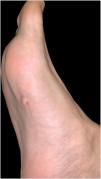
Physical Examination

Physical examination revealed a flesh-colored pedunculated tumor measuring 8mm in diameter. The tumor was soft, with a smooth surface, and was located on the middle third of the right sole (Fig. 1).

Figure 1
(0.06MB).
Histopathology

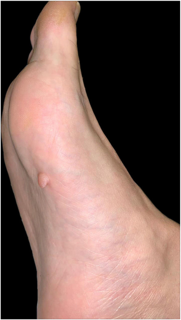
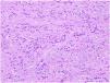
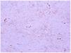
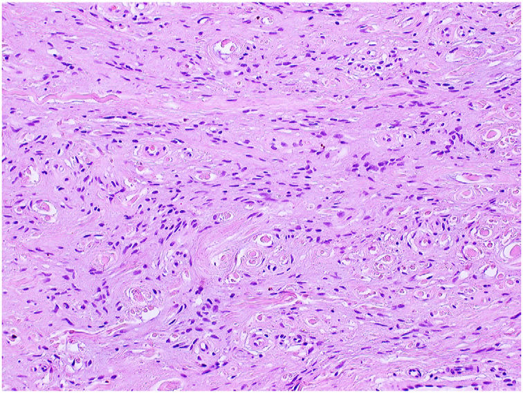
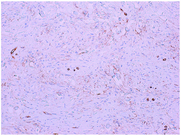
The tumor was removed using excisional biopsy. Histopathology revealed a proliferation of spindle-shaped homogeneous cells immersed in a myxoid stroma in the dermis (Fig. 2, hematoxylin–eosin, 200×). The results of immunohistochemistry were positive for CD34 and negative for S100 (Fig. 3, immunohistochemistry CD34, 200×).

Figure 2.

Hematoxylin–eosin, 200×.

(0.18MB).
Figure 3.

Immunohistochemical staining for CD34, 200×.

(0.15MB).

What is your diagnosis?

Diagnosis

Superficial acral fibromyxoma.

Clinical Course and Treatment

After diagnosis, the surgical margins were extended by 0.5mm. Histology revealed no relevant findings. No signs of recurrence have been observed after 10 months of follow-up.

Commentary

Superficial acral fibromyxoma is a benign mesenchymal tumor that mainly affects men in the fifth decade of life. In most published cases, the lesion is found in the periungual and subungual regions.1–3

Clinically, the tumor is usually nonencapsulated, well-circumscribed, polypoid or verrucous, and dome-shaped. It extends throughout the dermis, potentially as far as the subcutaneous tissue, underlying fascia, or periosteal layer.2–4

Radiology findings are nonspecific, although bone erosions or lytic lesions may co-occur in one-third of cases.1–5

Histopathology may reveal hyperkeratosis in the epidermis. Proliferation of spindle-shaped fibroblasts within a myxoid matrix, with nuclear atypia and mast cell infiltration, is a key finding in the dermis. Immunohistochemistry is positive for CD34, CD99, vimentin, and epithelial membrane antigen and negative for neural and muscular differentiation markers (S100, HMB-45, SMA, desmin, and actin).1,3,5

The differential diagnosis includes fibrokeratoma, fibroma, and plantar warts.3 It is also important to bear in mind tumors that are negative for CD34 (glomus tumor, sclerosing perineurioma, acquired digital fibrokeratoma, and periungual fibroma), as well as those that are positive for CD34 (superficial angiomyxoma, dermatofibrosarcoma protuberans, and myxoid neurofibroma).1,3,5

The treatment of choice is surgical resection with free margins, since local recurrence is recorded in approximately 20%–25% of cases. While the natural course of the tumor is benign, the histopathologic spectrum of spindle-shaped fibroblasts is variable, ranging from mild nuclear atypia to major atypia, with transformation to a low-grade malignant tumor being theoretically possible.4,6 However, prognosis is excellent, and, to date, there have been no published reports of malignant transformation or metastases.1,3–5

Conflicts of Interest

The authors declare that they have no financial or personal relationship with other persons or organizations that could give rise to a conflict of interest with respect to the present article.

References
[1]
E. Kalloniati, S. Arampatzi, C. Ouzouni, D. Papathemeli, E. Lazaridou, M.-G. Trakatelli.
Superficial acral fibromyxoma, appearing as retronychia: a rare clinical entity.
Skin Appendage Disord, 6 (2020), pp. 312-314
[2]
J.L. Sawaya, A. Khachemoune.
Superficial acral fibromyxoma.
Int J Dermatol, 54 (2015), pp. 499-508
[3]
V. Campbell, N. Machnikowski, J. Houghton, B. Murphy, O. Kerr.
Rare plantar heel presentation of superficial acral fibromyxoma.
Dermatol Ther, 33 (2020),
[4]
B.E. Crepaldi, R.D. Soares, F.D. Silveira, R.I. Taira, M.H. Matsumoto.
Superficial acral fibromyxoma in unusual location: a case report.
ANZ J Surg, 90 (2020), pp. 888-889
[5]
L. Crestani, I.A. Fasciani, P. Kakizaki, N.Y.S. Valente.
Case for diagnosis. Single-digit clubbing.
An Bras Dermatol, 95 (2020), pp. 524-526
[6]
J.F. Fetsch, W.B. Laskin, M. Miettinen.
Superficial acral fibromyxoma: a clinicopathologic and immunohistochemical analysis of 37 cases of a distinctive soft tissue tumor with a predilection for the fingers and toes.
Human Pathol, 32 (2001), pp. 704-714
Descargar PDF
Idiomas
Actas Dermo-Sifiliográficas
Opciones de artículo
Herramientas
es en

¿Es usted profesional sanitario apto para prescribir o dispensar medicamentos?

Are you a health professional able to prescribe or dispense drugs?