
Infiltration with botulinum toxin (BT) is considered an effective and widely used technique for treating plantar1 and palmar1,2 hyperhidrosis. Due to the high degree of sensory innervation—which depends on the posterior tibial, superficial peroneal, deep peroneal, saphenous, and sural nerves—pain is the main limiting factor. Therefore, performing a peripheral ankle block is essential, and it may also be used in other procedures, such as in tumor, dermatological, or other surgical conditions.
Description of the techniquePrior to the anesthetic block, antiseptic should be applied at the level of the ankle. For the blockade of the nerves described below, we use 2% lidocaine with a 21G intramuscular needle. Although the anesthetic block of the ankle can be performed under ultrasound guidance,3 a thorough knowledge of the anatomy may be sufficient to perform this technique safely and effectively.
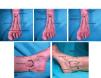
The posterior tibial nerve originates from the terminal branches of the sciatic nerve as they pass through the popliteal fossa. It runs deep along the midline of the posterior aspect of the leg, becoming superficial in the posterior region of the medial malleolus. It collects sensory afferents from the heel and the sole, except for 2 small medial and lateral areas3,4 (Fig. 1B). First, the posterior tibial artery is located at the upper end of the medial malleolus. Puncture is performed at the midpoint between the posterior edge of the upper malleolus and the medial border of the Achilles tendon. The injection is administered posterolateral to the artery until it reaches the periosteum (1–3cm). The needle is withdrawn 4–5mm, aspirated thoroughly, and 5–10mL of anesthetic solution is injected4 (Fig. 2D). As this nerve innervates most of the foot plantar surface, blocking this nerve alone may be sufficient for BT infiltration.

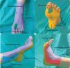
Sensory innervation of the posterior tibial (yellow), deep peroneal (green), superficial peroneal (magenta), saphenous (blue), and sural (red) nerves. (A) Dorsal aspect of the left foot. (B) Plantar aspect of the left foot. (C) Internal lateral aspect of the left foot. (D) External lateral aspect of the left foot.

The deep peroneal nerve originates from the common peroneal nerve. It emerges at the level of the fibular neck and travels deep to the extensor hallucis longus muscle. At the interosseous membrane, it becomes superficial, where it is lateral to the anterior tibial artery. It collects sensory afferents from the region between the first and second toe, the lateral side of the first toe, and the medial side of the second toe3,4 (Fig. 1A). By dorsiflexing the foot, the long extensor tendons of the toes and the great toe can be identified. The injection is administered at the inter-malleolar line, inserting the needle lateral to the dorsal artery of the foot and perpendicular to the skin. Once the bone has been reached, the needle is withdrawn a few millimeters, aspirated, and 5mL of anesthetic is injected4 (Fig. 2B).
The superficial peroneal nerve arises from the bifurcation of the common peroneal nerve, at fibular neck level, and descends along the lateral region of the leg. It provides sensation to the dorsum of the foot and the toes, except for a small area between the first and second toes3,4 (Fig. 1A). Starting from the injection point of the deep peroneal nerve, the needle is directed laterally toward the medial part of the lateral malleolus, and 5mL of anesthetic is injected in a fan shape4 (Fig. 2C).
The saphenous nerve originates from the terminal fibers of the femoral nerve. It is situated next to the great saphenous vein up to the medial malleolus. It provides sensation to the medial aspect of the leg, ankle, and heel3,4 (Fig. 1C). Starting from the injection point of the deep peroneal nerve, the needle is directed medially toward the upper and anterior area of the medial malleolus, and 3–5mL of anesthetic is injected4 (Fig. 2A).
The sural nerve arises from branches of the tibial and common peroneal nerves in the upper third of the calf. At the ankle, it is located in the posterolateral region, in contact with the small saphenous vein and lateral to the Achilles tendon. It collects sensory afferents from the lateral aspect of the heel, the proximal lateral third of the foot, and the lateral aspect of the fifth toe3,4 (Fig. 1D). The injection point is located between the upper end of the lateral malleolus and the Achilles tendon, posterior to the small saphenous vein. The needle is introduced from the outer border of the tendon and 5mL of local anesthetic is infiltrated subcutaneously up to the posterior aspect of the lateral malleolus4 (Fig. 2A). The duration of the anesthetic block depends on the anesthetic used. When using 2% lidocaine, the effect lasts from 45 to 60min. In general, after blocking the sensory afferents, it is advisable to use a cane to support ambulation for 2–6h after the block, due to the inability to adjust motor strength to sensory feedback.4
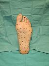
Technique for botulinum toxin infiltrationThe Minor's test can be used to detect the areas most affected by hyperhidrosis. To prepare the BT dilution, for each vial of 100 units of type A BT (Botox, Allergan, Irvine, CA, USA), 5mL of 0.9% physiological saline is added. Between 100 and 200 units are injected per sole, depending on the extent and size. The injection is administered at the dermis/subcutaneous junction, using a sterile 30G needle, spacing the points 1–2cm apart, for a total of 15–50 points (Fig. 3). At each point, 0.1cm3 (2 units) is injected. The anticholinergic effect begins in 7–10 days and lasts for a maximum of 6–10 months.5

The indications, contraindications, and complications of the technique are similar to those for the palmar region.2
In summary, infiltration with BT using a peripheral ankle block is a safe and effective procedure, especially when an ultrasound device is not available.
Conflicts of interestNone declared.


