

No studies focused on counting the interdigital acquired melanocytic nevi (AMN) of the foot (IDNf) have ever been conducted. Therefore, our objective was to study the relationship between the presence of IDNf and the total number of AMN in the feet and the rest of the body, the racial phenotypic characteristics, and other risk factors for melanoma.
Material and methodsWe conducted a cross-sectional observational study with 255 patients ≥18 years old who attended our Dermatology Unit from September 2020 through February 2021, and included all AMN ≥1mm from the feet and ≥2mm from the rest of the bod. The association between the variables was studied using univariate and multivariate logistic regression models.
ResultsThe presence of IDNf was significantly and independently associated with the presence of plantar AMN and body counts ≥50 AMN. However, no significant differences were observed regarding sex, age, personal history of melanoma, presence of nevi on the dorsum of the foot, history of sunburn or UV rays, or racial phenotypic characteristics.
ConclusionsThe presence of IDNf is associated with a higher count of plantar nevi and total AMN in the body, meaning that interdigital spaces of the foot – anatomical expansions of the sole and other possibly genetic causes – could be responsible for the number of AMN found in this location, as these regions are not photoexposed.
Existe una ausencia de estudios centrados en el recuento de nevus melanocíticos adquiridos (NMA) interdigitales de los pies (NIDp). Nuestro objetivo fue estudiar la relación de la presencia de NIDp con el número total de NMA de los pies y del resto del cuerpo, las características fenotípicas raciales y otros factores de riesgo de melanoma.
Material y métodosSe realizó un estudio observacional transversal que incluyó a 255 pacientes, ≥18 años, que acudieron a nuestro servicio de Dermatología desde septiembre de 2020 a febrero de 2021. Se registraron todos los NMA ≥1mm de los pies y ≥2mm del resto del cuerpo. Se estudió la asociación entre las variables mediante un modelo de regresión logística uni- y multivariante.
ResultadosLa presencia de NIDp se asoció de manera estadísticamente significativa e independiente con la presencia de NMA plantares y con recuentos de ≥50 NMA en el cuerpo. Sin embargo, no se observaron diferencias significativas respecto al sexo, la edad, los antecedentes personales de melanoma, la presencia de nevus en el dorso de los pies, antecedentes de quemaduras solares o rayos UVA, ni con características fenotípicas raciales.
ConclusionesLa presencia de NIDp se relaciona con un mayor recuento de nevus plantares y NMA totales del cuerpo, lo que puede significar que los espacios interdigitales son una expansión anatómica de la planta del pie y que otras causas, posiblemente genéticas, serían responsables del número de NMA en esta localización, al ser zonas nada fotoexpuestas.

Acquired melanocytic nevi (AMN) are benign tumors of melanocytes considered the most common benign neoplasms in the white population.1 It is well known that the number of AMN is one of the most important risk factors for developing melanoma.2
Although most AMN are usually located in sun-exposed (SE) areas, they also appear in less-exposed sites such as the palms or soles, and these are known as acral AMN.3 Publications reporting counts of these AMN are quite scarce.3–13 Their pathogenesis is unknown, and they display a different mutational profile vs non-acral nevi and acral lentiginous melanoma (ALM).14 On the other hand, the possible association between ALM and acral AMN is controversial.13,15,16
However, there are no studies characterizing AMN in the interdigital spaces, an area especially hidden from SE. Although this location is a transitional zone between the dorsum and the sole of the feet, it is unclear whether the presence of AMN in interdigital spaces can be equated to those located in either of these two areas, or whether there is any relationship with the number of nevi on the rest of the body.
The aim of the present study was to observe whether the count of interdigital nevi of the feet (IDNf) was associated with the total number of AMN on the foot, and other risk factors for developing melanoma, such as the total number of AMN of the body (AMN-B), racial phenotypic characteristics, and a history of UV radiation (UVR) exposure.15
Material and methodsWe designed a cross-sectional observational study including all adult patients (≥18 years) who attended the pigmented lesion clinic of the Dermatology Department at Hospital General Universitario de Alicante (GUHA) from September 2020 through February 2021. Patients with Fitzpatrick skin types I to IV5 were selected, and those meeting any of the following exclusion criteria were excluded: presence of giant congenital nevi, severe mental illness or poor hygiene conditions that prevented examination of the feet.
The study protocol was conducted in full compliance with the principles of the Declaration of Helsinki and approved by the GUHA Ethics Committee (Act 2021-03).
Study variablesThe outcome variable was the number of interdigital AMN ≥1mm on the feet.
The following explanatory variables were considered: sex, age, eye color (light [blue or green] and dark [brown or black]); hair color at age 20 (light [red, blond, or light brown], dark [dark brown or black]); skin phototype (light [I and II] or dark [III and IV]), presence of freckles, personal history of severe sunburn (defined as blistering after sun exposure), personal history of melanoma, non-melanoma skin cancer (NMSC), and non-skin cancer, use of UVA lamp, number of AMN-B ≥2mm (≤50 and >50), number of AMN ≥1mm on the sole (AMN-S) and dorsum of the feet (AMN-D), referencing those underneath the malleoli.
Regarding the cutoff point for considering acral AMN, including IDNf, we chose ≥1mm as the minimum size to avoid confusion with other lesions, especially solar lentigines on the dorsum. For the cutoff in AMN-B, we chose ≥2mm, as this is widely established16,17 and our group had experience from a previous study.18
Data collection method and study variablesData collection was performed in two phases. The first phase consisted of an interview, where inclusion and exclusion criteria were reviewed. In the second phase, patients were examined in their underwear to count AMN-B and those on the feet, including interdigital spaces and excluding areas covered by underwear and the scalp. All patients were assigned an identification number to ensure anonymity. A dermoscopic examination of all suspicious lesions was performed.
All data were collected by a previously trained dermatology resident (V.S.G.), supervised by an expert dermatologist in pigmented lesions (J.B.).
Statistical analysisNormality of quantitative variables (age and number of AMN) was assessed using the Kolmogorov–Smirnov test. Since the number of AMN-D and AMN-S did not follow a normal distribution, the median and interquartile range [IQR] were used. The Mann–Whitney U test was used to study the relationship between qualitative and quantitative variables. Lastly, the Spearman correlation test was used to assess the relationship between quantitative variables.
Categorical variables were expressed as absolute and relative frequencies. Continuous variables were categorized for subsequent statistical analysis: age was divided into tertiles and the AMN of the feet (dorsum, sole, and interdigital) as present or absent. Binary logistic regression was used in the bivariate analysis. To quantify the size of association, the odds ratio (OR) with a 95% confidence interval (95% CI) was used. Statistically significant associations were further analyzed in a multivariate analysis, calculating the adjusted OR (aOR), with a 95% CI. Additionally, an analysis of the crude number of IDNf and the number of AMN-D and AMN-S was performed.
The level of significance for hypothesis testing was set at p<0.05. Statistical analysis was performed using SPSS software (version 25.0, IBM Corp, Armonk, NY, USA).
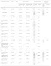
ResultsOf the 257 subjects invited to participate in the study, 2 (0.8%) were excluded due to poor foot hygiene. Finally, a total of 255 patients who met the selection criteria were included, 51.4% of whom were women, with a median age of 53 years (Table 1).
Descriptive, bivariate, and multivariate analysis of the patient sample (n=255).
| Explanatory variables | Total(n=255) | Result variables | Bivariate analysis | Multivariate analysis | |||
|---|---|---|---|---|---|---|---|
| Interdigital AMN (yes)=44 | Interdigital AMN (no)=211 | OR (95% CI) | p-Value* | aOR (95% CI) | p-Value | ||
| Age, n (%) | |||||||
| <45 years | 90 (35.6) | 25 (56.8) | 65 (31.1) | 4.2 (1.7–10.4) | 0.002 | 0.8 (0.3–2.5) | 0.71 |
| 45–63 years | 79 (31.2) | 12 (27.3) | 67 (32.1) | 2.0 (0.7–5.3) | 0.179 | 1.8 (0.6–5.2) | 0.30 |
| >63 years | 84 (33.2) | 7 (15.9) | 77 (36.8) | 1 | – | 1 | – |
| Sex (male), n (%) | 124 (48.6) | 19 (43.2) | 105 (49.8) | 1.3 (0.7–2.5) | 0.43 | ||
| Eye color (light), n (%) | 91 (35.7) | 16 (36.4) | 75 (35.5) | 1 (0.5–1.9) | 0.92 | ||
| Hair color (light), n (%) | 98 (38.4) | 16 (36.4) | 82 (38.9) | 1.1 (0.6–2.2) | 0.76 | ||
| Phototype (light), n (%) | 35 (13.7) | 6 (13.6) | 29 (13.7) | 1.0 (0.4–2.6) | 0.99 | ||
| Freckles (presence), n (%) | 93 (36.5) | 20 (45.5) | 73 (34.6) | 1.5 (0.8–3.0) | 0.21 | ||
| History of sunburn (yes), n (%) | 137 (53.7) | 24 (54.5) | 113 (53.8) | 1.0 (0.5–2.0) | 0.95 | ||
| UVA exposure, n (%) | 31 (12.2) | 5 (12.3) | 26 (11.4) | 1.1 (0.4–3.0) | 0.90 | ||
| AMN body (>50), n (%) | 137 (53.7) | 34 (77.3) | 103 (48.8) | 3.6 (1.7–7.6) | 0.001 | 3.5 (1.5–7.9) | 0.003 |
| AMN dorsum of the foot, median (p25–p75) | 0 (0–1) | 2 (1–2) | 1 (1–2) | 0.11 | |||
| AMN sole of the foot, median (p25–p75) | 0 (0–1) | 2 (1–2) | 1 (1–2) | <0.001 | |||
| Presence of AMN dorsum, n (%) | 118 (46.3) | 24 (54.5) | 94 (44.5) | 1.5 (0.8–22.9) | 0.23 | ||
| Presence of AMN sole, n (%) | 99 (38.8) | 28 (63.6) | 71 (33.6) | 3.5 (1.8–6.8) | <0.001 | 2.4 (1.2–5.1) | 0.020 |
| Personal history of NMSC (yes), n (%) | 51 (20.0) | 3 (6.8) | 48 (22.7) | 0.3 (0.1–0.8) | 0.025 | 0.3 (0.8–1.2) | 0.09 |
| Personal history of MM (yes), n (%) | 61 (23.9) | 6 (13.6) | 55 (26.1) | 0.5 (0.2–1.1) | 0.09 | 0.5 (0.2–1.3) | 0.15 |
| History of other cancers, n (%)a | 20 (7.9) | 4 (9.1) | 16 (7.6) | 1.2 (0.4–3.8) | 0.74 | ||
Statistically significant results in bold.
NMSC: non-melanoma skin cancer; CI: confidence interval; AMN: acquired melanocytic nevi; MM: malignant melanoma; OR: crude odds ratio; aOR: adjusted odds ratio; UVA: ultraviolet A.
Types of cancer: breast carcinoma (n=7), prostate carcinoma (n=2), Hodgkin's lymphoma (n=2), vulvar carcinoma (n=1), colon carcinoma (n=1), acute leukemia (n=1), liposarcoma (n=1), lung carcinoma (n=1), rectal carcinoma (n=1), renal carcinoma (n=1), urinary tract carcinoma (n=1), bladder carcinoma (n=1).
Table 1 presents the differences in explanatory variables based on the presence or absence of IDNf and the results of the bivariate analysis. The crude analysis showed that only patients <45 years were significantly associated with the presence of IDNf. On the other hand, no significant differences were observed regarding sex, eye color, hair color, phototype, presence of freckles, history of sunburn, UVR exposure, personal history of melanoma, or non-skin cancer. However, a personal history of NMSC showed a negative or “protective” association with the presence of IDNf.
The relationship of the presence of IDNf with variables on the number of nevi was as follows: an association was observed with the presence and number of AMN-S. Conversely, no association was observed with the presence or number of AMN-D. Similarly, a high number of AMN-B (≥50 AMN) was associated with the presence of IDNf.
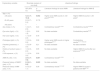
Furthermore, we observed that the AMN-S count increased significantly with the IDNf count, with a correlation coefficient of 0.248 (p<0.001) (Table 2). By contrast, there was no significant correlation between the counts of AMN-D and IDNf.
Correlation study between the number of interdigital nevi (outcome variable) and the number of nevi on the sole and dorsum of the feet (explanatory variables).
| Explanatory variable | Result variable | |
|---|---|---|
| No. of interdigital nevi | ||
| r | p-Value | |
| No. of plantar AMN | 0.248 | <0.001 |
| No. of dorsal AMN | 0.105 | 0.095 |
Statistically significant results are shown in bold.
After multivariate analysis, the variables independently associated with IDNf were the presence of AMN-S (aOR, 2.4; 95% CI, 1.2–5.1; p=0.020) and the presence of >50 AMN-B (aOR, 3.5; 95% CI, 1.5–7.9; p=0.003) (Table 1).
DiscussionThis study demonstrated that the presence of IDNf is related to the number of AMN-S, and AMN-B.
The number of skin AMN is a known risk factor for cutaneous melanoma2 and has been associated with racial phenotypic traits and SE.19–21 Counts of acral AMN, areas minimally exposed to UVR, have rarely been reported in the literature and, when present, almost exclusively refer to the soles and palms, with the dorsum not being studied.3–13 Furthermore, the interdigital spaces are even more concealed.
Finally, a total of 255 patients were included, of whom 17.3% presented IDNf. Regarding age, we observed that patients younger than 45 years had a higher risk of having IDNf. There are no studies on counts of IDNf, so the information we have is indirect from AMN-B. It has been reported that acral AMN increase during adolescence to middle age and decrease in older age.5,22 Palicka and Rhodes9 and Kogushi-Nishi et al.13 found higher prevalences of acral AMN in individuals younger than 50 years (Table 3). Similarly, the number of AMN-B follows a similar pattern.1 Although there are no specific studies on IDNf counts, in our study the highest risk of having IDNf was observed in patients younger than 45 years.
Comparison of the bivariate analysis results from our study with findings from the literature.
| Explanatory variable | Bivariate analysis of our study | Literature findings | ||
|---|---|---|---|---|
| OR (95% CI) | p-Value* | Literature findings for acral AMN | Literature findings for AMN-B | |
| Age, n (%) | ||||
| <45 years | 4.2 (1.7–10.4) | 0.002 | Higher acral AMN counts in <50 years5,9,13,22 | Higher AMN-B counts in <50 years1 |
| 45–63 years | 2.0 (0.7–5.3) | 0.179 | ||
| >63 years | 1 | – | ||
| Sex (male), n (%) | 1.3 (0.7–2.5) | 0.43 | Contradictory results9,10,23 | Contradictory results10,24,25 |
| Eye color (light), n (%) | 1 (0.5–1.9) | 0.92 | No data available | Contradictory results23,26 |
| Hair color (light), n (%) | 1.1 (0.6–2.2) | 0.76 | ||
| Phototype (light), n (%) | 1.0 (0.4–2.6) | 0.99 | Higher acral AMN counts in higher phototypes5,9,11 | Contradictory results10,17,19,20,23 |
| Freckles (presence), n (%) | 1.5 (0.8–3.0) | 0.21 | No data available | No data available |
| History of sunburn (yes), n (%) | 1.0 (0.5–2.0) | 0.95 | No data available | Higher AMN-B counts after sun exposure21,23,24 |
| UVA exposure, n (%) | 1.1 (0.4–3.0) | 0.90 | ||
| AMN-B (>50), n (%) | 3.6 (1.7–7.6) | 0.001 | Contradictory results9,10,24 | – |
| Personal history of NMSC (yes), n (%) | 0.3 (0.1–0.8) | 0.025 | No data available | No data available |
| Personal history of MM (yes), n (%) | 0.5 (0.2–1.1) | 0.09 | Contradictory results13,19,22 | Higher AMN-B counts in patients with MM27,28 |
Statistically significant results are shown in bold.
NMSC: non-melanoma skin cancer; CI: confidence interval; AMN: acquired melanocytic nevi; AMN-B: acquired melanocytic nevi of the body; MM: malignant melanoma; SE: sun exposure; OR: crude odds ratio; aOR: adjusted odds ratio; UVA: ultraviolet A.
We found no significant differences in the presence of IDNf by sex. Regarding acral nevi, Palicka and Rhodes9 found higher prevalences of acral AMN in white women vs men, while other authors found no differences at all.10,23 As for the number of AMN-B, results are inconsistent, with some studies finding higher counts in men,24 others in women,25 and some finding no sex difference.10
We did not observe any relationship between IDNf counts and eye or hair color. No publications were found relating the number of acral AMN with these features. Regarding the number of AMN-B, results are inconsistent: some authors found no relationship,26 while others concluded that subjects with light eyes and hair have more AMN.23
We found no differences either with respect to the presence of IDNf and phototype or presence of freckles. As for counts of acral nevi and skin color, some authors observed that individuals with darker skin have more palmar nevi than whites, with no differences for AMN-S,5,11 while others found that the prevalence of palmar and plantar nevi increases with pigmentation.9 Regarding AMN-B counts, although some studies found more AMN in fair-skinned than in dark-skinned subjects,17,19,20,23 others found no relationship.10,24,25
We did not observe an association between IDNf and UVR exposure. As interdigital spaces are particularly covered from SE, and as AMN usually appear in sun-exposed areas,1 this lack of association seems logical. Although different patterns of ALM have been described according to location and relative sun exposure,26 nothing has been published on acral AMN in this regard. In general, it has been described that AMN-B increase after SE.21,23,24
The presence of IDNf was significantly associated with a higher risk of elevated AMN-B count. Counts of acral AMN have been associated with higher AMN-B counts,9,24 although other studies have not found this association.10 As for the relationship with acral AMN count, bivariate analysis showed that the presence of IDNf was associated with a greater risk of having an AMN-S, whereas IDNf were not related to AMN-D.
The presence of IDNf was associated with a lower risk of having NMSC. This protective role could be explained by the fact that IDNf, being in covered areas (unlike most NMSC), have different pathogenic factors. On the other hand, we found no reports in the literature linking acral nevus counts and NMSC.
Another notable finding is that the presence of IDNf was not associated with a personal history of melanoma. The relationship between the number of AMN-S and the past medical history of melanoma varies in the literature. Some studies found no difference in the prevalence of AMN-S between plantar melanoma cases and controls13; others have associated ALM with high counts of AMN-S,16 and others concluded that AMN count is an important risk factor for non-acral melanoma, but not for ALM.22 In general, it has been demonstrated that high AMN-B counts are associated with a high risk of melanoma.27,28
Finally, in multivariate analysis, the only two variables that independently explained the presence of IDNf were the presence of AMN-S and the presence of >50 AMN on the body. We hypothesize that this association between IDNf and plantar nevi may be due to the anatomical similarity of interdigital spaces to the soles, making them an extension of the sole. The absence of relationship with AMN-D suggests that other anatomical, genetic, or even photoexposure factors may influence the development of AMN-D, but not AMN-S or IDNf.
The relationship between site-specific nevus count and AMN-B has been described several times.29 The best correlations have been found for the number of nevi on the forearm and thigh. We found no studies specifically addressing IDNf. However, Papageorgiou et al.10 detected a relationship between the presence of acral nevi and elevated AMN-B counts, consistent with our findings between IDNf and AMN-B.
On the other hand, acral AMN and ALM do not seem to have a direct relationship.30 The two entities have independent clinical and histopathological characteristics, and a different mutational profile, suggesting that ALM arise mostly de novo, not from AMN.
Regarding the examination of interdigital spaces, we think it is not routinely performed in daily practice, which could delay the diagnosis of malignant disease. In fact, in our study we detected one case of in situ interdigital ALM the patient was unaware of.31
As for the study limitations, it is a single-center study with a small sample size, which limits its external validity.
In conclusion, the presence of IDNf is associated with a higher number of AMN-S and AMN-B. As far as we know, this is the first published study on IDNf, providing a new epidemiological perspective on acral nevi in an area of the body without SE, such as interdigital spaces. Furthermore, the association we observed between the presence of IDNf and the number of AMN-B is possibly due to genetic factors, and not only environmental factors such as SE, influencing the number of AMN.
FundingNone declared.
Conflicts of interestNone declared.


