


The patient was an 82-year-old woman with no history of interest who consulted for an asymptomatic lesion that had first appeared 20 years previously on the supraclavicular region. The lesion had remained stable over time.
Physical ExaminationThe lesion was nodular, elastic, well delimited, and flesh-colored with a violaceous-bluish tone. It was not adherent to deeper planes (Fig. 1).
Additional Tests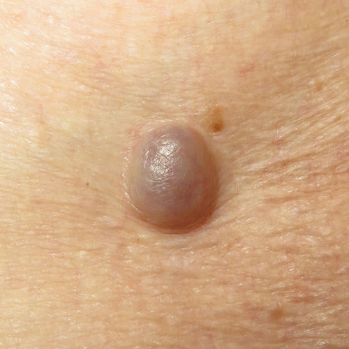
B-mode ultrasound with a linear 22MHz probe (MyLabTM Gamma Esaote) revealed a well-delimited dermal-hypodermal lesion with 2 clearly differentiated areas (Fig. 2). The first was a superficial, spherical, hypoechoic area with mobile hyperechoic structures in the interior that ran from the superior to the inferior pole to create a “snow falling” image (Video 1 of the Additional material). The second area was deeper, solid, heterogeneously echoic, and richly vascularized in color Doppler.
Histopathology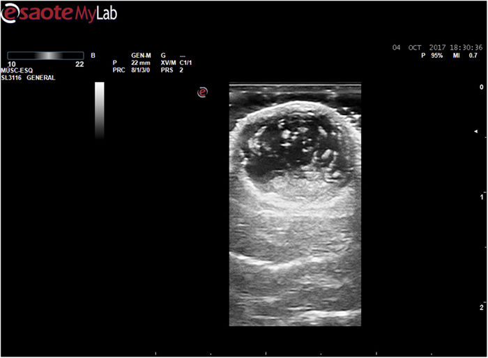
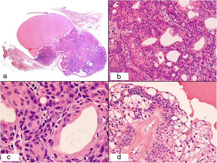
The lesion was removed. Histopathology revealed a nonencapsulated well-delimited tumor (Fig. 3) with a large cystic component covered by a flat epithelium and a solid area. The solid area comprised 2 cell populations. The first cells had eosinophilic cytoplasm with oval or vesicular nuclei and prominent nucleoli (Fig. 3C). The second cell population comprised smaller cells with clear cytoplasm and small and eccentric nuclei (Fig. 3D). Apocrine decapitation secretion was visible in some areas, as were areas of ductal and squamous differentiation (Fig. 3B) and mucinous metaplasia in keloid-like stroma.
What is your diagnosis?
DiagnosisApocrine hidradenoma.
ProgressThe lesion was removed completely, with no recurrence after 1 year of follow-up.
CommentApocrine hidradenoma, which is also known as clear cell hidradenoma, solid-cystic hidradenoma, nodular hidradenoma, and eccrine acrospiroma, is an uncommon benign tumor. Clinical diagnosis is often difficult owing to its variable clinical presentation and requires a high degree of suspicion. Therefore, the lesion generally has to be removed and undergo histology analysis in order to confirm the diagnosis.
The ultrasound characteristics of apocrine hidradenoma have received little attention in the literature.1–5 The most commonly reported characteristic is the presence of a double solid-cystic component that is richly vascularized in Doppler color mode. It is important to remember that as some forms of apocrine hidradenoma are exclusively solid, other possible ultrasound findings should also be taken into consideration.
The presence of a fluid-fluid level and the snow falling sign, which are specific to this entity, were recently described.5
In the “fluid-fluid” pattern, we can see a cystic cavity with a clear level between a superior more hypoechoic area (less dense) and an inferior hyperechoic area (more dense). This finding highlights the presence of at least 2 fluids with different echodensity. While this finding is common in various fields of ultrasound imaging (eg, complicated hydroceles with hemorrhage or ovarian teratoma),6 it has not been reported in other dermatological conditions.
In the snow falling sign, we can see mobile echogenic structures falling from the superior to the inferior pole within the tumor. The origin of these structures is not clear. A similar movement can be seen in some cases of hydrocele where the content is heterogeneous. The content of the cystic cavity in apocrine hidradenoma has not been established, although, in order to explain this phenomenon, we can postulate that it must be heterogeneous. The various theories on the origin of this heterogeneity include bleeding within the cystic cavity, detritus from the epithelium lining the cavity, or even the apocrine secretion found in some cells of this tumor.
We report a new case of apocrine hidradenoma with ultrasound findings. We need studies that shed light on the composition of the cystic component of these tumors, as well as a description of the ultrasound findings in other variants of the condition.
Conflicts of InterestThe authors declare that they have no conflicts of interest.
We are grateful to Drs. José Luis Díaz Recuero and Alejandra Pérez Plaza for their help in the preparation of the article and to Dr. Luis Requena Caballero for his willingness and interest in teaching.
Please cite this article as: Toro EMd, González YCP, Cembranos MDM. Tumor de larga evolución con patrón «en copos de nieve»Eczema y urticaria en Portugal. 2019;110:765–766.


