
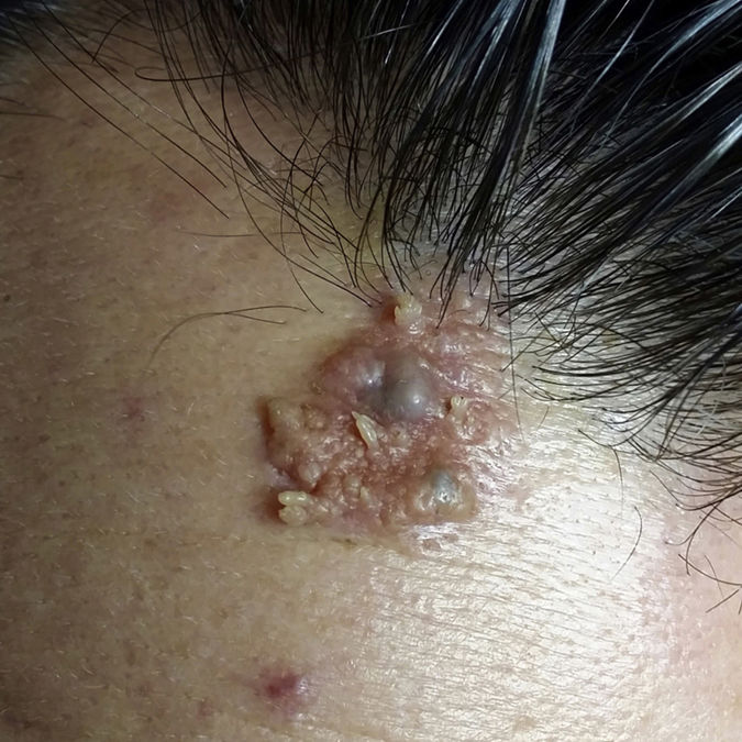
A 38-year-old man presented with a waxy congenital plaque on his forehead that had undergone changes in the past few years. These changes included the development of several bluish nodules within the plaque. The texture of these nodules had also varied. Physical examination showed a well-delimited yellowish plaque measuring 3cm containing 2 blue nodules and surrounded by several warty, papillomatous areas (Fig. 1).

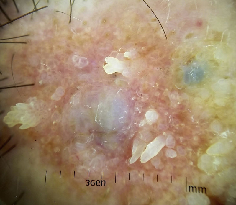
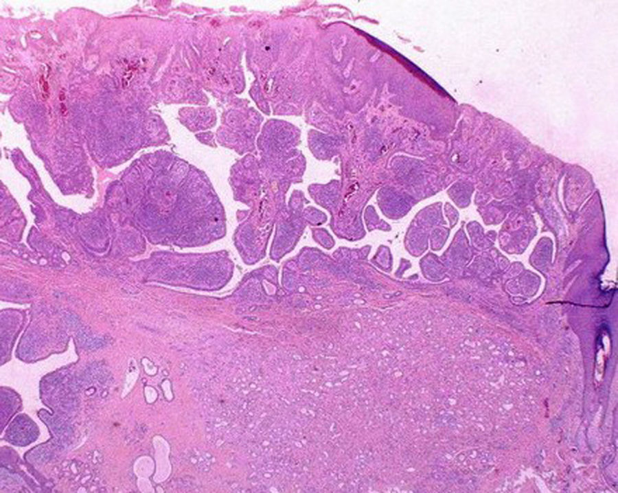
Dermoscopic examination showed a white-yellowish cobblestone pattern with 2 large, asymmetric blue-gray ovoid nests, the largest of which contained thick vessels. It also showed a symmetric erythematous lesion with exophytic papillary structures and polymorphous (dotted and linear) vessels (Fig. 2). The histologic images are shown in Figure 3.

.")
Diagnosis: Collision tumors or tumors composed of trichoblastoma and syringocystadenoma papilliferum on a sebaceous nevus.
Sebaceous nevus is a benign congenital hamartoma that usually affects the scalp or face.1 It is well known that secondary tumors with a predominant line of differentiation (follicular, sebaceous, or apocrine) can arise in sebaceous nevi. These tumors are observed in approximately 10% to 20% of cases. The 2 main secondary tumors associated with sebaceous nevus are trichoblastoma and syringocystadenocarcinoma papilliferum, which are generally benign. Malignant tumors can also develop but they are rare. The most common type is basal cell carcinoma. Few studies have described the dermoscopic features of lesions arising in sebaceous nevus. Zaballos et al.2 described the main characteristics of these tumors in a review of 58 cases. Basal cell carcinomas tend to appear as large, blue-gray, asymmetric ovoid nests possibly containing blue-gray globules. Trichoblastomas, by contrast, tend to have more symmetric, homogeneous ovoid nests occupying the entire lesion. Basal cell carcinomas tend to start with characteristic arborizing telangiectasias. Trichoblastoma overlying a sebaceous nevus has also been described as an irregular blue-gray area with a linear vascular pattern or arborizing telangiectasias with white structures.3 Syringocystadenocarcinoma papilliferum shows a symmetric pattern formed by an exophytic papillary structure with erythema, ulceration, and atypical vessels (coma, hairpin, horseshoe-shaped, and atypical irregular, glomerular, dotted, and polymorphous) vessels.4 On occasions, this erythematous background may be divided by whitish linear structures that demarcate lobules containing different types of vessels.5 Apocrine hidrocystomas appear as symmetric homogeneous areas with arborizing telangiectasias.
Dermoscopy is one of the most accurate in vivo tools available for guiding diagnosis prior to surgery. It is also useful for monitoring disease progression and enabling the early detection of tumors arising in sebaceous nevi. More descriptive studies of the characteristics of these tumors are needed to aid diagnosis. Histologic examination, however, provides the greatest accuracy and is the gold standard diagnostic tool, as the entities can sometimes mimic each other on dermoscopy.6
Conflicts of InterestThe authors declare that they have no conflicts of interest.
Please cite this article as: Lobato-Berezo A, Aguilera-Peiró P, Pujol RM. Tumores de colisión sobre nevus sebáceo: claves para su diagnóstico dermatoscópico. Actas Dermosifiliogr. 2018;109:647–648.


