
Pagetoid dyskeratosis is a frequent incidental finding in various skin lesions.1
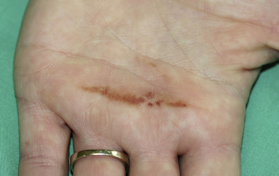
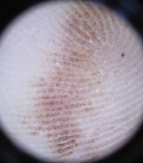
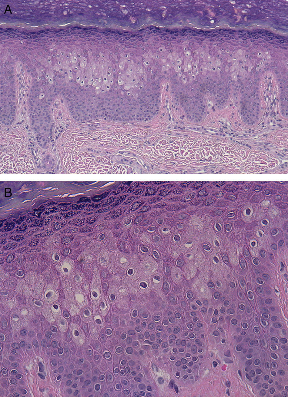
The patient was a 27-year-old woman who consulted for an asymptomatic hyperpigmented lesion that had arisen on the left palm 18 months earlier. Examination revealed a dark brown, hyperpigmented macule measuring 40×3mm, with well-defined borders and a linear appearance (Fig. 1). Dermoscopy showed a parallel pattern of the cutaneous ridges (Fig. 2). Incisional biopsy of the lesion revealed dyskeratotic cells in a pagetoid pattern (Fig. 3). Based on these findings we made a diagnosis of pagetoid dyskeratosis of the palm. Cryotherapy led to complete resolution of the lesion. After 2 years the patient remains practically asymptomatic. A very faint yellow macule persists at the same site, with no significant alterations on dermoscopy.


Pagetoid dyskeratosis can present clinically as a hyperpigmented macule. A literature search revealed only 3 cases in which this condition affected the palm.2–4 To date, the dermoscopic pattern of a lesion located on the hand has only been described in 1 case.2 That patient presented a dark brown macule over the distal phalanx of the fifth finger; the dermoscopic features included a parallel pattern of the cutaneous ridges and a fibrillary pattern that led to the need to exclude palmoplantar melanoma.
The etiology and pathogenesis of this condition are unknown, although it appears to be related to recurrent friction over the area. The prognosis is good.
On histopathology, dyskeratotic cells are observed together with larger-than-usual, round keratinocytes with pale cytoplasm and a pyknotic nucleus surrounded by a pale halo, in a pagetoid distribution.1
The dermoscopy findings in this patient should be noted, as this pattern was suggestive of malignant melanoma, though it was also compatible with a diagnosis of subcorneal hematoma. Pagetoid dyskeratosis must be included in the differential diagnosis of pigmented palmar lesions that show a parallel cutaneous ridge pattern on dermoscopy.
We would like to thank Dr. Luis Requena for his help in reaching the diagnosis of the lesion in this patient.
Please cite this article as: Loidi L, Mitxelena J, Córdoba A, Yanguas I. Dermatoscopia de la disqueratosis pagetoide palmar. Actas Dermosifiliogr. 2014;105:804–805.


