


In-transit melanoma metastases are defined as deposits of tumor cells in the cutaneous and/or subcutaneous planes occurring between the primary melanoma and the regional lymph node. They may be present at diagnosis or develop as recurrences in 5–10% of patients, and their presence has been associated with an adverse prognosis. The complexity and heterogeneity of this condition, together with the lack of studies specifically focused on the affected population, make it difficult to establish evidence-based treatment algorithms. This review aims to describe, from a practical perspective, the therapeutic options available for patients with in-transit melanoma metastases, including local, regional, and systemic treatments.
Melanoma is an aggressive neoplasm and the leading cause of death from skin cancer, despite accounting for only 1% of these cancers.1–3 Five-year survival depends on disease extent and is >99% for localized disease, 73% for regional involvement, and 35% for distant metastases.4
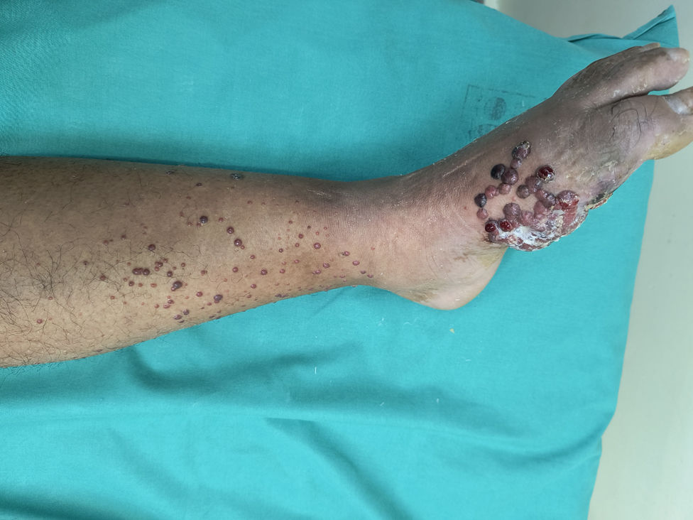
In-transit melanoma (ITM) refers to cutaneous or subcutaneous deposits of tumor cells located between the primary tumor and the regional lymph node and has been reported in 5–10% of cases. Classically, ITMs are defined as clinically evident accumulations of tumor cells located >2cm from the primary tumor, “satellites” when located within the surrounding 2cm, and “microsatellites” when neoplastic cells are diagnosed microscopically (Fig. 1).1 These three entities exhibit similar biological behavior and are associated with a poor prognosis2; therefore, they are classified as stage III according to the American Joint Committee on Cancer staging system (AJCC-8).5

Clinical photograph of a patient presenting with multiple erythematous and blackish papules and nodules on the lower extremity, corresponding to “satellite” metastases (within 2cm of the primary tumor) and “in-transit” metastases (>2cm from the primary tumor toward the corresponding lymph node).
The mechanisms underlying the development of ITM are not fully understood; trapping of tumor cells within dermal lymphatic vessels has been proposed, among several other mechanisms. Recent studies suggest that ITMs harbor specific mutations, such as Q61 mutations in the NRAS gene, and have a lower mutational burden, which could explain their locoregional behavior.7–9
Moreover, distinct risk factors have been described for lymphatic and hematogenous dissemination. Age, primary tumor location, tumor thickness, and vascular invasion have been associated with lymphatic recurrence; in contrast, tumor thickness, absence of regression, the TERT promoter, and BRAF mutation have been associated with hematogenous metastasis.3
Risk factors for the development of ITM include a positive sentinel lymph node, ulceration,6,12 Breslow thickness >2mm, and location on the lower extremities, head/neck, or acral sites.4 Therefore, ITMs differ from hematogenous metastases in their biological behavior, prognosis, and treatment.8,10,11
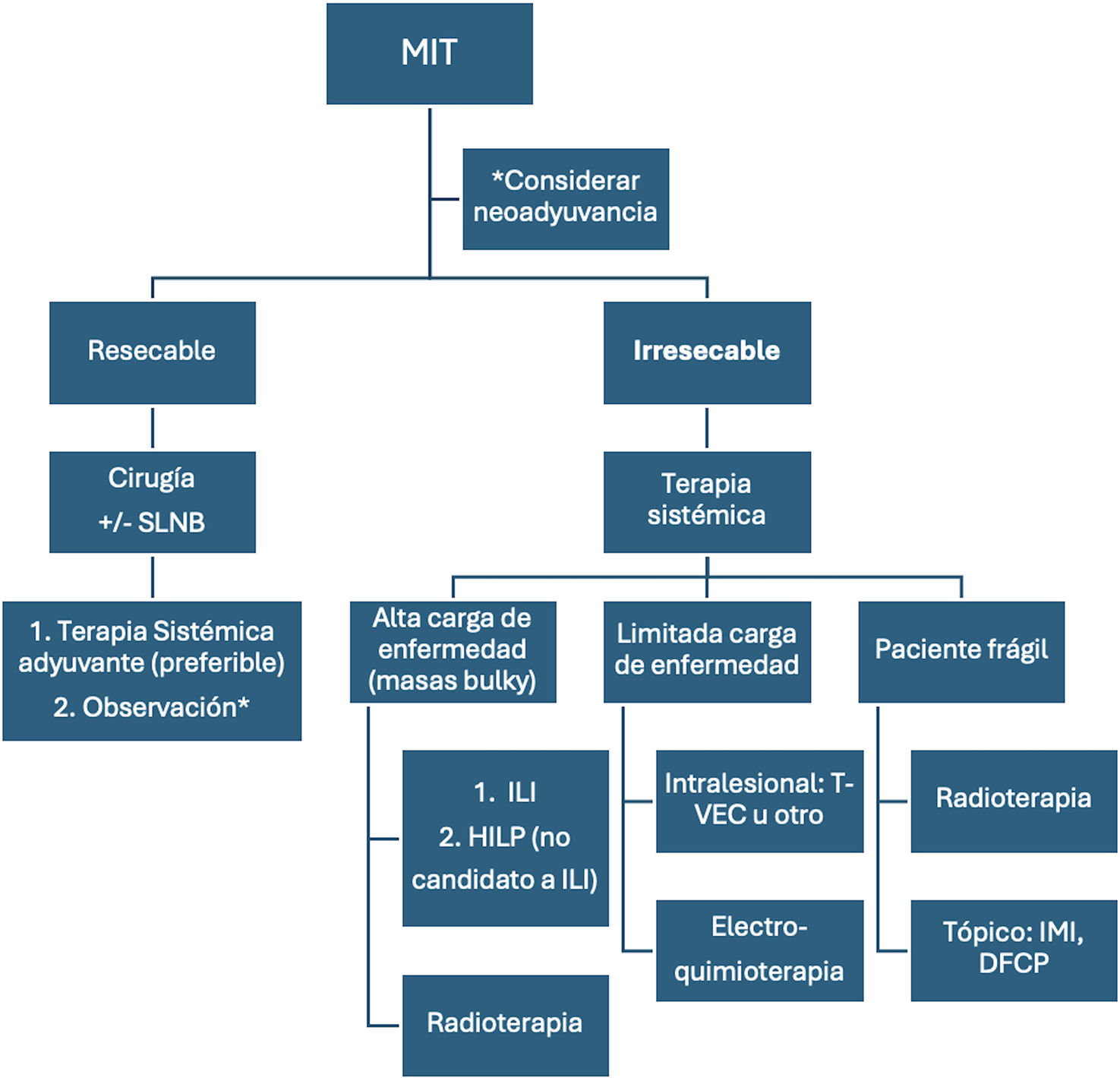
Because of its complexity and the limited available evidence, treatment should be tailored to the clinical context. This review addresses therapeutic options for ITM without lymph node involvement, including surgery and regional and systemic therapies, taking into consideration factors such as tumor burden, distant disease, and the patient's performance status (Fig. 2).6,8,10,11

Algorithm of therapeutic alternatives for in-transit melanoma. ITM, in-transit metastasis; SLNB, sentinel lymph node biopsy; HILP, hyperthermic isolated limb perfusion; ILI, isolated limb infusion; IMI, imiquimod; DPCP, diphencyprone. *The decision between observation and adjuvant therapy should take into consideration the risk of melanoma recurrence, treatment toxicity, the disease-free interval, and distant metastasis-free survival.
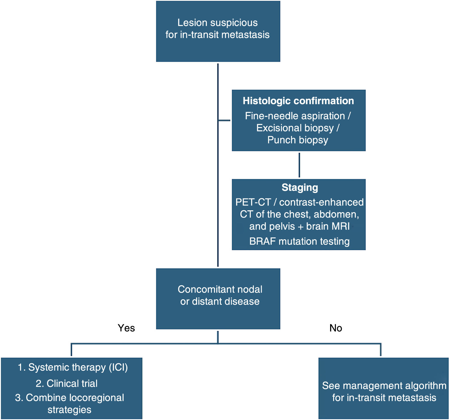
In a patient with a past medical history of melanoma, any cutaneous or subcutaneous lesion, whether pigmented or nonpigmented, particularly if ipsilateral to or close to the primary lesion, should raise suspicion of ITM.5 The surrounding soft tissues and regional lymph node basin should be palpated.5 Histopathological assessment of the suspicious lesion is performed by fine-needle aspiration, excisional biopsy, or incisional biopsy (Fig. 3).10,13

A total of 75% of patients with ITM will have regional or distant involvement6; therefore, once the diagnosis is confirmed, staging should be repeated using brain magnetic resonance imaging and PET/CT (if PET/CT is unavailable, contrast-enhanced computed tomography may be performed).5,7,8 Testing for BRAF mutations is recommended,7,8 regardless of the mutational profile of the primary tumor because of the possibility of discordant BRAF mutation status (Fig. 3).9
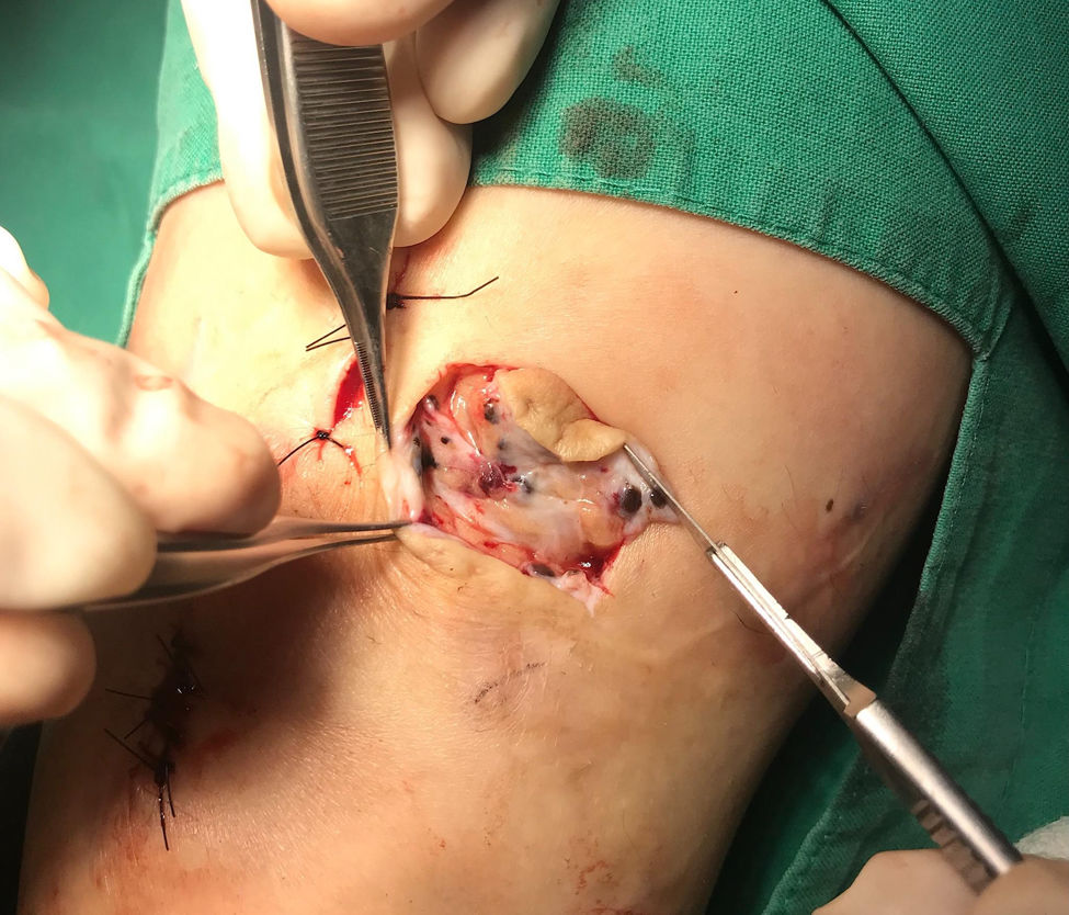
Excision of ITM and role of sentinel lymph node biopsyIn patients with a low burden of resectable disease, surgical excision is the standard of care.7,8 Lesions are considered resectable if they can be removed with negative margins without significantly compromising the function or aesthetics of the affected area.10 Some groups consider disease resectable when there are no more than three lesions and none is >5cm (Fig. 4).5 This definition does not take into consideration the anatomical region involved or the patient's ability to tolerate surgery.

There is no evidence supporting a specific excision margin. The National Comprehensive Cancer Network (NCCN) and European Society for Medical Oncology (ESMO) clinical practice guidelines recommend excision with clear margins,8 whereas other groups have suggested 1-cm margins.11 The disadvantage of surgical excision is that it does not eliminate underlying metastatic cells, particularly those located along lymphatic vessels (Fig. 4). Nevertheless, surgery may improve the patient's quality of life.
The role of sentinel lymph node biopsy (SLNB) in the setting of ITM is controversial and should be discussed by a multidisciplinary tumor board. Given its technical feasibility, SLNB is suggested when the result would alter the patient's therapeutic options or provide additional prognostic information.7,8
In patients with a positive SLNB, there is no evidence of a survival benefit from lymphadenectomy,2,12 but these patients are candidates for adjuvant immunotherapy.2,8
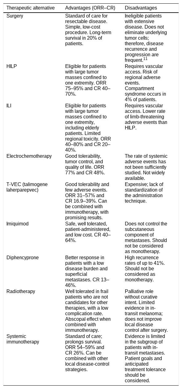
Regional therapyWhen disease is unresectable because of its extent, distribution, or anatomical involvement, regional therapies allow treatment of the disease site with minimal risk of systemic adverse events, delaying progression and reducing symptoms.13 Furthermore, they may complement systemic therapies (Table 1).
Available therapies for in-transit melanoma metastases.
| Therapeutic alternative | Advantages (ORR–CR) | Disadvantages |
|---|---|---|
| Surgery | Standard of care for resectable disease. Simple, low-cost procedure. Long-term survival in 20% of patients. | Ineligible patients with extensive disease. Does not eliminate underlying tumor cells; therefore, disease recurrence and progression are frequent.11 |
| HILP | Eligible for patients with large tumor masses confined to one extremity. ORR 75–95% and CR 40–70%. | Requires vascular access. Risk of regional adverse events. Compartment syndrome occurs in 4% of patients. |
| ILI | Eligible for patients with large tumor masses confined to one extremity, including elderly patients. Limited regional toxicity. ORR 40–80% and CR 20–40%. | Requires vascular access. Lower rate of limb-threatening adverse events than HILP. |
| Electrochemotherapy | Good tolerability, tumor control, and quality of life. ORR 77% and CR 48%. | The rate of systemic adverse events has not been sufficiently studied. Not widely available. |
| T-VEC (talimogene laherparepvec) | Good tolerability and few adverse events. ORR 31–57% and CR 16.9–39%. Can be combined with immunotherapy, with promising results. | Expensive; lack of standardization of the administration technique. |
| Imiquimod | Safe, well tolerated, patient-administered, and low cost. CR 40–64%. | Does not control the subcutaneous component of metastases. Should not be considered as monotherapy. |
| Diphencyprone | Better response in patients with a low disease burden and superficial metastases. CR 13–46%. | High recurrence rates of up to 41%. Should not be considered as monotherapy. |
| Radiotherapy | Well tolerated in frail patients who are not candidates for other therapies, with a low complication rate. Abscopal effect when combined with immunotherapy. | Palliative role without curative intent. Limited evidence in in-transit melanoma; does not improve local disease control after surgery. |
| Systemic immunotherapy | Standard of care; prolongs survival. ORR 54–59% and CR 26%. Can be combined with other local disease-control strategies. | Evidence is limited in the subgroup of patients with in-transit metastases. Patient goals and anticipated treatment tolerance should be considered. |
ORR, overall response rate; CR, complete response.
Regional chemotherapy is indicated when lesions are confined to one extremity. Although it is used less frequently because of the success of systemic therapies, it has demonstrated good local disease control and may therefore be considered as an adjunct to systemic therapy if the patient has progressed during systemic treatment or is ineligible for systemic therapy.7,8,14 The principle underlying these techniques is the administration of chemotherapy at doses up to 20 times higher than those tolerated systemically. They require vascular access and isolation of the affected extremity.5 The two techniques are hyperthermic isolated limb perfusion (HILP) and isolated limb infusion (ILI).8
Hyperthermic isolated limb perfusion (HILP)This technique involves direct dissection and cannulation of the major vessels of the affected extremity, isolation using a tourniquet, followed by hyperthermia of the extremity (39–41°C), and finally melphalan infusion.15 An overall response rate (ORR) of 75–95% and a complete response (CR) rate of 40–70% have been reported.15 Although addition of tumor necrosis factor alpha to HILP produced CR rates of up to 70%,16,17 these findings were not reproducible.18 The procedure carries a high risk of severe regional adverse events, including a 4% incidence of compartment syndrome.5,15
Isolated limb infusion (ILI)ILI follows the same principle as HILP but uses less invasive venous access and has several advantages. It does not require specialized equipment, is well tolerated even by patients older than 80 years, has low systemic toxicity and limited limb toxicity, and is considerably less expensive than systemic therapies.19 ORRs of 40–80% and CR rates of 20–40% have been reported in ITM14; consequently, it has become the first option for patients who are ineligible for systemic or injectable treatments.14 Greater effectiveness has been demonstrated when it is used early in patients with a low tumor burden.14
Both techniques have similar rates of regional toxicity, and no significant differences have been found in time to disease progression or survival.5 The advantage of ILI is its lower rate of severe limb-threatening regional adverse events; therefore, HILP is reserved for patients with large tumor masses who are ineligible for ILI.15 In contrast, HILP provides higher CR rates and greater long-term effectiveness.5,15
ElectrochemotherapyElectrochemotherapy consists of the coadministration of a poorly permeable cytotoxic drug (bleomycin or cisplatin) along with short local electrical pulses. This increases cell membrane permeability and allows the drug to diffuse into the cells.20 The chemotherapeutic agent may be administered intravenously or intratumorally, with similar effectiveness.21
An ORR of 77% and CR rate of 48% have been reported,21 and evidence supports its use in combination with systemic therapies.22 It is included in the ESMO guideline for the management of unresectable ITM because of its good tolerability, tumor control, and improvement in quality of life.7
Intralesional therapyIntratumoral administration is considered when ITMs are numerous and unresectable. The main limitations are the lack of standardization of the administration technique and limited availability within health care services.13
Although many agents have been studied without demonstrating an overall survival benefit,13 viral oncolytic therapies have shown promising results.23 Their mechanism of action involves selective infection of tumor cells with oncolytic viruses, thereby stimulating the host antitumor immune response through preferential viral replication within tumor cells. This represents a favorable strategy even in patients with a poor response to immune checkpoint inhibitors.23,24
Talimogene laherparepvec (T-VEC) is the only agent approved by the U.S. Food and Drug Administration (FDA) for the management of unresectable melanoma.25 It is based on modified human herpes simplex virus type 1 (HSV-1).23 It demonstrated good tolerability and long-term efficacy, with an ORR of 31% and a CR rate of 16.9%, without a significant survival benefit.26 Other studies have reported an ORR of 57% and a CR rate of 39%;27 accordingly, it is included among the NCCN clinical guideline recommendations.8 Another HSV-1 derivative is vusolimogene oderparepvec (RP1), with preliminary results showing an ORR of 62.5% when combined with nivolumab in patients naïve to systemic therapy and an ORR of 36% in patients who failed to respond to anti-PD-1 and anti-PD-1/anti-CTLA-4 therapy.28 CVA21, derived from coxsackievirus A21, has demonstrated ORR efficacy similar to that of T-VEC.23 Other viruses, including reovirus, adenovirus, and poxvirus, have not demonstrated favorable responses.23
Cytokines are another intralesional option. IL-2 increases tumor immunogenicity but is poorly tolerated when administered systemically at effective doses.29 Intralesional administration has produced a CR rate of 69%, with no response at uninjected sites and no effect on survival; in addition, the frequency of administration (twice weekly) and the high rate of local adverse events should be considered.30 Darleukin tavokinogene telseplasmid (tavo) is a plasmid encoding IL-12 that increases local expression of IL-12 and interferon. It has shown modest results, with a CR rate of 18% and an ORR of 36%31; when combined with immunotherapy, an ORR of 30% has been reported, along with responses at uninjected sites.32
One of the major advantages of intratumoral therapy is its ability to be combined with systemic agents (see below); consequently, numerous agents are currently under investigation (Daromun, ONCOS-102, BT-001, Canerpaturev, PVSRIPO, LL37, APX005M, etc.).13
Topical therapyTopical therapy is safe, well tolerated, patient-administered, and inexpensive.33 Imiquimod is a Toll-like receptor 7 and 8 agonist and has been used off-label for the treatment of ITM.11 The 5% formulation has been used in case series and small studies employing various treatment schedules. CR rates of up to 64.3% have been reported with imiquimod alone11 and 40% when combined with 5-fluorouracil.34 Furthermore, one case series reported a 65% response rate with cryosurgery followed by imiquimod, with CR achieved in 40% of responders.35
Diphencyprone (DPCP) is a contact immunosensitizer. It promotes selective destruction of tumor cells by lymphocytes while preserving unaffected tissue.36 Studies have used DPCP in an aqueous cream at concentrations of 0.005–1%, applied with a 1-cm margin once or twice weekly for 24–48h.36 CR rates of 13–46% have been demonstrated,33,36,37 with a significant survival benefit in patients achieving a complete response.36 Factors associated with clinical benefit included a low disease burden (<15 lesions)37 and epidermotropic morphology.36 Like imiquimod, DPCP does not control the subcutaneous component of metastases and therefore has high recurrence rates of up to 41%; moreover, failure to achieve sensitization has been found to reflect a high disease burden.37 It should be considered in patients receiving palliative care and never as monotherapy.11
RadiotherapyThe evidence regarding radiotherapy (RT) for ITM is controversial.38 A retrospective study of patients with ITM showed that postoperative RT did not improve local disease control.38 Conversely, its role in unresectable disease has not been studied, although it may reduce tumor burden and alleviate pain and bleeding.39 In addition, it is associated with a low rate and severity of complications,40,41 making it a palliative alternative in elderly patients who are ineligible for more aggressive therapies.
More recently, the abscopal effect has been described, namely, a response occurring at a site distant from the irradiated area during immunotherapy (see combination therapies).39,42
Systemic therapiesSystemic therapy with immune checkpoint inhibitors (ICIs) or targeted therapy is the standard treatment for stage III and IV melanoma.7,8 In particular, anti-CTLA-4 (ipilimumab), anti-PD-1 (pembrolizumab/nivolumab), and anti-PD-L1 therapies have demonstrated exceptional clinical efficacy,43,44 providing the basis for the development of standard treatments for ITM. However, response to immunotherapy is not universal45; therefore, pretreatment molecular markers are essential for patient stratification, improving efficacy, and reducing treatment-induced toxicity.45
Although clinical trials have included patients with ITM, evidence in this subgroup remains limited. Retrospective studies have evaluated responses to different immunotherapy regimens in patients with ITM, with encouraging results.46,47 The first study showed an ORR of 54%, a CR rate of 26%, and a 2-year survival rate of 63%, with anti-PD-1 monotherapy achieving the best results (CR, 56%), followed by combined anti-PD-1/anti-CTLA-4 therapy (ORR, 40%) and anti-CTLA-4 therapy (ORR, 38%).46 Another study reported similar results, with an ORR of 63% and a 36-month survival rate of 80%; anti-PD-1 monotherapy achieved an ORR of 59%.47
Overall, the available evidence suggests that adjuvant immunotherapy extends survival in patients with ITM. The ESMO guideline recommends systemic therapy (preferably anti-PD-1) for patients with resectable ITM and a short disease-free interval (<6 months) or for those with unresectable ITM.7 The NCCN guideline proposes systemic therapy as first-line therapy for ITM.8 Both clinical practice guidelines indicate that observation vs immunotherapy may be considered according to the patient's goals and anticipated treatment tolerance.7,8
Regarding neoadjuvant therapy, most clinical trials either did not include patients with satellite or in-transit lesions or included only a small number of such lesions.48 Nevertheless, given the high degree of activity observed in other stage III/IV settings,48 the NCCN guideline recommends consideration of neoadjuvant therapy.8
Combination therapyMultiple ongoing clinical trials are evaluating combinations of systemic and regional therapies.13 For example, combined T-VEC has been associated with significantly longer survival and progression-free survival (PFS) than T-VEC monotherapy.23,49 ILI followed by ipilimumab produced an ORR of 85%, a CR rate of 62%, and 1-year PFS of 58%.50 DPFP combined with nivolumab has shown favorable results in several case reports,51,52 among other approaches.
Finally, synergy between RT and ICIs has recently been identified. The immune escape mechanism of tumor cells involves upregulation of immunosuppressive ligands and cytokines within the tumor microenvironment, thereby reducing antigen presentation. RT may reverse this process through downregulation of CD47, increased MHC-1 expression on tumor cells, and promotion of immune-cell migration into the tumor, among other mechanisms.53,54 This systemic effect may induce an abscopal response in distant tumors.39,53 Therefore, combining immunotherapy with RT may maximize therapeutic efficacy in primary and metastatic melanoma.39 However, the optimal dose and fractionation schedule remain to be determined, and toxicity associated with different ICI combinations requires further evaluation.39
ConclusionsITM is a therapeutic challenge requiring multidisciplinary and individualized management. Systemic therapies have significantly improved prognosis and are the standard of care for advanced disease. In patients with a low tumor burden, surgery combined with adjuvant immunotherapy may be curative. In patients with unresectable or rapidly growing lesions, local therapies represent a well-tolerated option that can be combined with systemic therapies, improving prognosis and quality of life.
Conflict of interestThe authors declare no conflict of interest.