


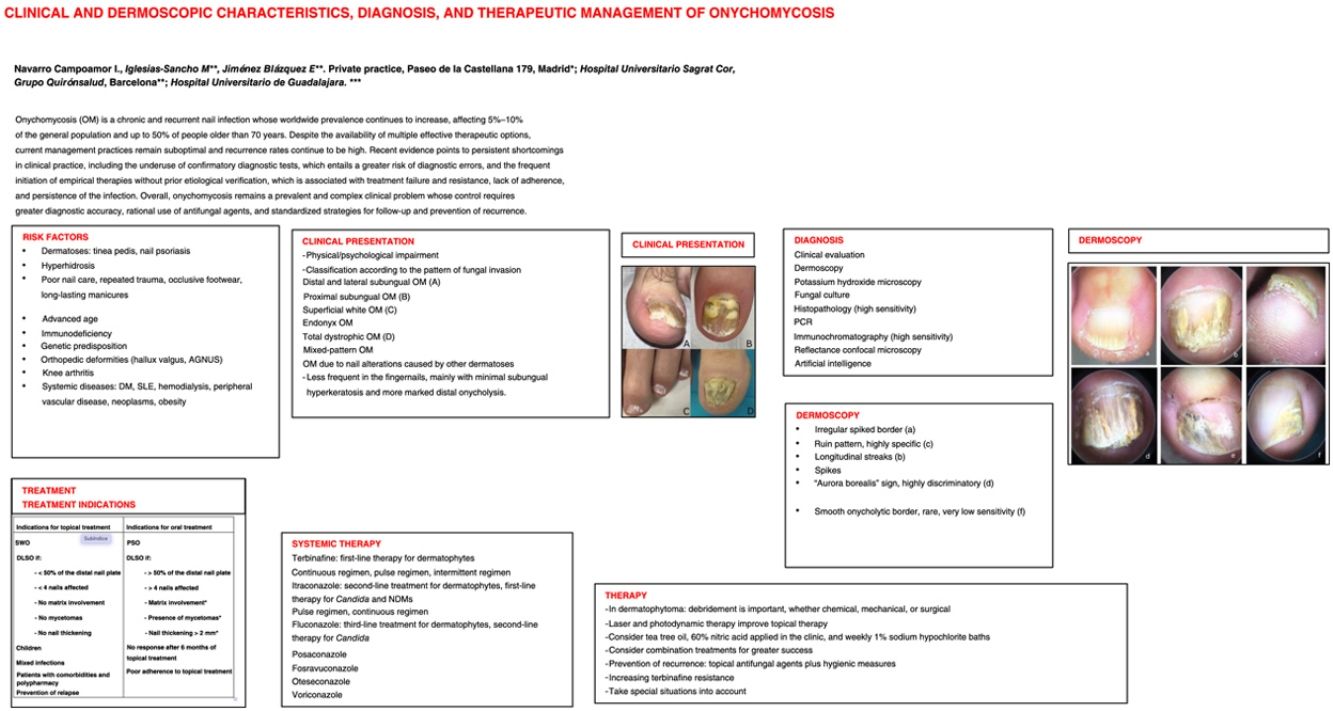
Onychomycosis is a chronic and recurrent nail infection whose worldwide prevalence continues to increase, affecting 5–10% of the general population and up to 50% of individuals older than 70 years. Despite the availability of multiple effective therapeutic options, current management practices remain suboptimal, and recurrence rates continue to be high.
Recent evidence points to persistent shortcomings in clinical practice, including the underuse of confirmatory diagnostic tests, which entails a greater risk of diagnostic errors, and the frequent initiation of empirical treatment without prior etiological verification, which is associated with treatment failure and resistance, lack of adherence, and persistence of infection.
Overall, onychomycosis remains a prevalent and complex clinical problem whose control requires greater diagnostic accuracy, rational use of antifungal agents, and standardized strategies for follow-up and prevention of recurrence.
La onicomicosis es una infección ungueal crónica y recurrente cuya prevalencia mundial continúa en aumento, afectando entre el 5–10% de la población general y hasta al 50% de las personas mayores de 70 años. Pese a la disponibilidad de múltiples opciones terapéuticas eficaces, las prácticas actuales de manejo siguen siendo subóptimas y las tasas de recurrencia continúan siendo elevadas.
La evidencia reciente señala deficiencias persistentes en la práctica clínica, entre ellas la infrautilización de pruebas diagnósticas confirmatorias, que conlleva un mayor riesgo de errores diagnósticos, y la frecuente instauración de tratamientos empíricos sin verificación etiológica previa, lo que se asocia a fallos terapéuticos y resistencias, falta de adherencia y persistencia de la infección.
En conjunto, la onicomicosis continúa siendo un problema clínico prevalente y complejo, cuyo control exige una mayor precisión diagnóstica, uso racional de antifúngicos y estrategias uniformes de seguimiento y prevención de recurrencias.

Onychomycosis (OM) is a chronic and recurrent nail disease, with an estimated worldwide prevalence of between 5.5% and 10%, which increases with age and affects up to 50% of people older than 70 years, while being uncommon in the pediatric population.1–3 It is the most common nail disorder, accounting for 50–60% of nail dystrophies, with a predominance in the toenails.2,4,5
Its chronic course is related to diagnostic delay, as it is frequently underestimated by patients as a merely cosmetic problem. Despite successful initial treatment, relapse or reinfection rates reach 20–25%, and overall recurrences may be as high as 53%, reflecting its persistence and therapeutic difficulty.4,6
Clinically, it manifests as nail thickening, chromonychia, onycholysis, and, occasionally, periungual involvement. Its high prevalence and functional and cosmetic impact make it a relevant health problem, with a significant effect on quality of life, particularly in women and in patients with fingernail involvement.7
Prevalence and etiology vary according to age, climate, and habits. Dermatophyte infections, termed tinea unguium, account for 60–90% of cases; non-dermatophyte molds (NDMs) for approximately 10%, occurring more frequently in women and individuals older than 60 years; and yeasts for 10–20%, mainly in fingernails because of exposure to moisture.1,4,8,9 Mixed infections, present in up to 11%, are usually more resistant and recurrent, and their clinical interpretation remains complex.1,4 In addition, the formation of fungal biofilms on the nail plate contributes to persistence and resistance to treatment.
Risk factorsRisk factors for OM include various dermatoses, particularly tinea pedis and nail psoriasis (NP), which presents concomitant OM in >50% of cases.10 In patients with NP, OM should be ruled out before initiating conventional or biologic treatments, given the higher incidence described with anti-TNF therapy.11 Hyperhidrosis and onychodystrophy of any origin are also predisposing factors.
Exogenous factors include poor nail care, repeated trauma—such as that associated with sports—, occlusive footwear, and long-lasting manicures.12
Advanced age is one of the most common risk factors, together with tinea pedis and repeated microtrauma. In children, OM is uncommon, although it may be more prevalent in Down syndrome13 or act as a marker of immunodeficiency. There is a genetic predisposition, particularly in chronic OM,14 and contact with affected family members increases the risk.
Similarly, foot deformities such as hallux valgus and Asymmetric Gait Nail Unit Syndrome (AGNUS) are associated, the latter with a greater predisposition to NDM infection.9 Greater susceptibility has recently been described in patients with knee arthritis, probably because of limited mobility and inadequate hygiene.2
Other associated systemic diseases include immunosuppression, diabetes mellitus, lupus erythematosus, hemodialysis, peripheral vascular disease, neoplasms, and obesity.
Clinical presentationOM typically manifests as chromonychia, nail thickening, subungual hyperkeratosis, and onycholysis.4,5 It affects patients both physically and psychologically, impairing their daily activities, causing stigmatization, and generating fear of contagion.16,17 Toenails are the most widely affected, particularly the great toenail.18
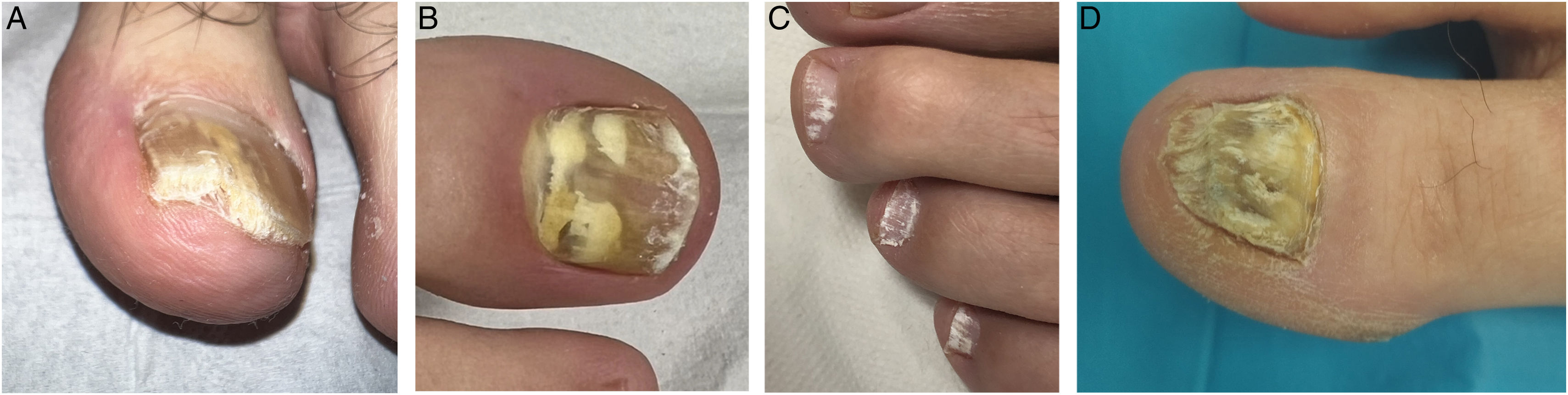
Hay and Baran classified OM in 2011 according to the pattern of fungal invasion, subsequently providing a practical clinical approach19,20 (Fig. 1):
- -
Distal and lateral subungual OM (DLSO): The most common clinical form; invasion begins in the distal and/or lateral subungual area, with subungual hyperkeratosis and secondary onycholysis. When DLSO is due to NDMs, it is usually associated with periungual erythema.9 The most frequently associated agents are Trichophyton rubrum, Trichophyton mentagrophytes, Epidermophyton floccosum, Fusarium, Scopulariopsis breviculatis, and Candida albicans.
- -
Proximal subungual OM (PSO): Uncommon; invasion occurs from the proximal nail area. In the absence of paronychia, it may be a marker of immunodeficiency; in the presence of paronychia, it is generally related to moisture or hyperhidrosis, where NDMs are more frequent—particularly Aspergillus spp., Fusarium spp., and Scopulariopsis brevicaulis—and Candida. Of note, the importance of nail examination in immunocompromised patients, in whom PSO caused by Fusarium spp. may represent the portal of entry for hematogenous dissemination.9 The most widely implicated dermatophyte fungus is T. rubrum.
- -
Superficial white OM (SWO): Rare. It is characterized by fungal invasion of the dorsal surface of the nail plate, producing white patches that are easily removed by scraping. The most widely implicated fungi are Trichophyton interdigitale and Fusariun spp. Occasionally, NDMs may give rise to “deep” SWO.9
- -
Endonyx OM (EO): A rare form in which the nail plate is directly invaded without onycholysis or subungual hyperkeratosis; milky-white patches and compact lamellar scaling are observed within the nail plate. It is more prevalent in Asia and Africa. The main etiological agents are Trichophyton soudanense and Trichophyton violaceum, although T. rubrum and others may be involved in isolated cases.21
- -
Total dystrophic OM (TDO): Rare. It is characterized by progressive and complete destruction of the nail plate. It may develop after prolonged progression of any OM subtype, particularly chronic DLSO, or occur as a primary form in infections caused by highly invasive dermatophytes. Management is complex. It may form part of an immunodeficiency state (as in chronic mucocutaneous candidiasis). It is often caused by dermatophytes such as T. rubrum or T. mentagrophytes.
- -
Mixed-pattern OM: Characterized by the presence of >2 subtypes. The most common coexistence in toenails is DLSO plus SWO.4
- -
OM due to nail alterations caused by other dermatoses.

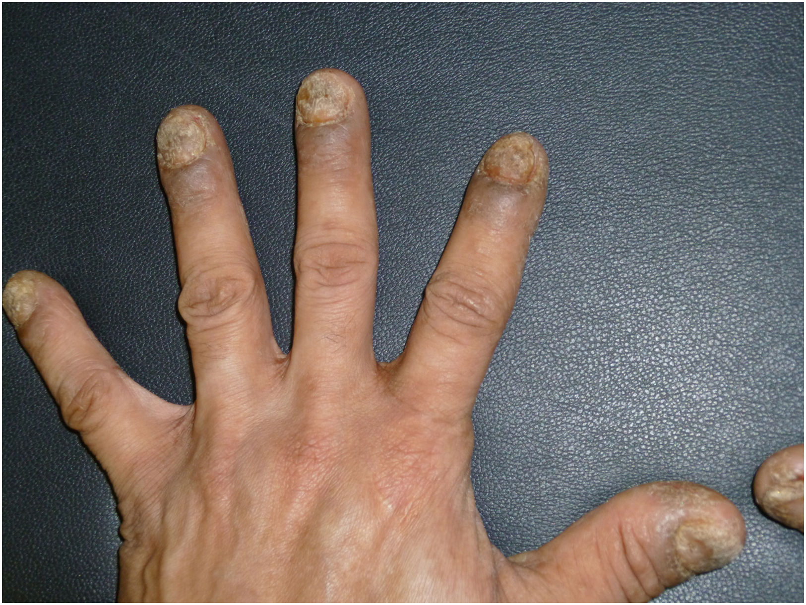
Fingernail OM is 7 times less frequent than toenail OM and has been poorly described. It predominates on the right hand, in the 2nd and 3rd fingers, and is associated with chronic moisture exposure, previous paronychia, and impairment of the nail barrier. It accounts for approximately 10–20% of cases; although the most frequent agents are Candida spp., followed by dermatophytes such as T. rubrum, recent studies indicate a predominance of dermatophytes over Candida.22 These authors describe it as presenting minimal subungual hyperkeratosis with more marked onycholysis and propose a clinical and onychoscopic classification into 4 subtypes:
- -
Chaotic pattern, with combinations of color, mild onycholysis, and paronychia.
- -
Hyperkeratotic subtype (the most common), with moderate-to-severe onycholysis and hyperkeratosis with little ruin-like appearance.
- -
Superficial involvement subtype.
- -
“Crumbling” subtype, with marked destruction of the nail plate (Fig. 2).

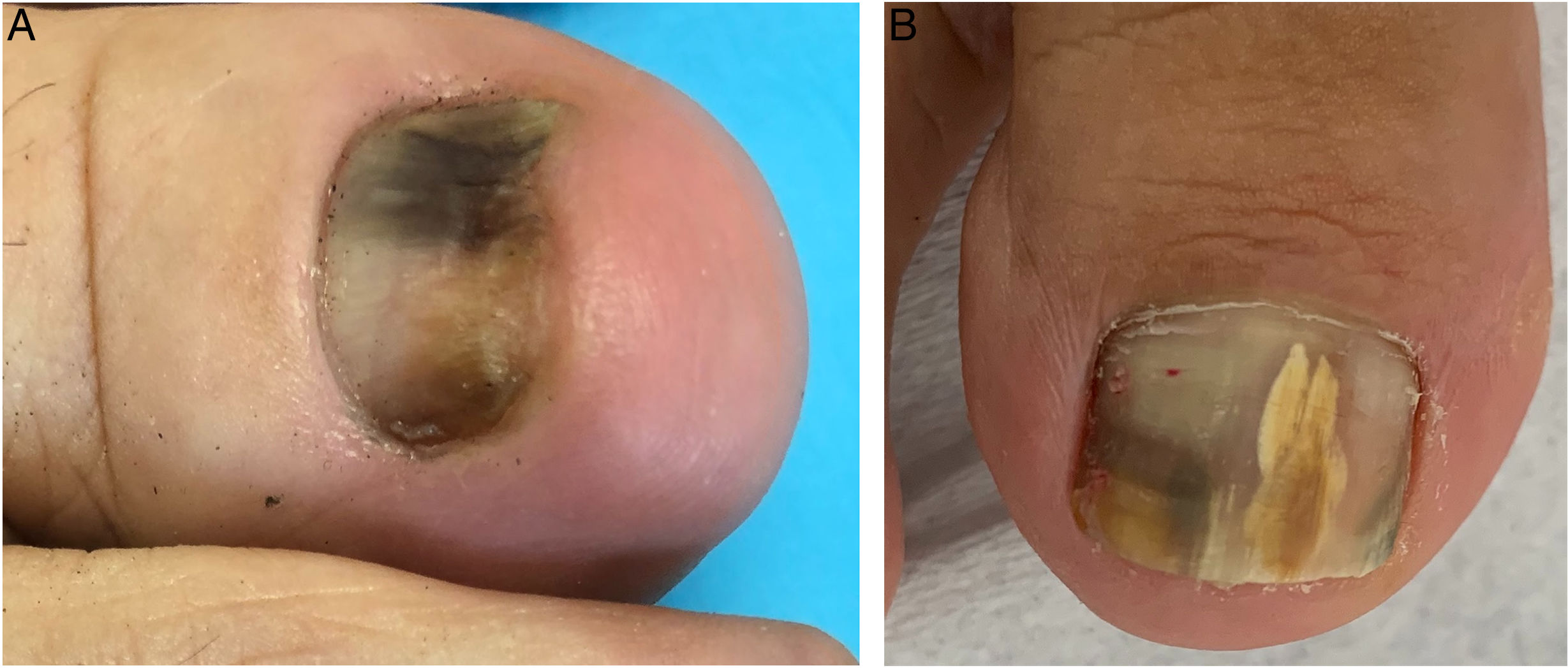
A rare form caused by fungal species capable of producing pigment themselves or by activation of melanocytes in the nail matrix. Although the main pathogen is T. rubrum, it may also be caused by NDMs such as Aspergillus niger, Neoscytalidium, Alternaria, Fusarium and, more rarely, Candida.23,24

Brownish or blackish nail discoloration is observed and may be linear, diffuse, or localized. The reverse triangular pattern, subungual hyperkeratosis, and coexistence with white or yellow lines may provide useful diagnostic clues.
Green nailOM predisposes to superinfection by Pseudomonas aeruginosa, which produces a greenish-black discoloration, although certain isolated fungal infections may produce it.25
DermatophytomasDermatophytomas are characterized by localized hyperkeratotic nail masses of dense fungal elements associated with biofilms, with marked refractoriness to oral antifungal therapy. Dermatophytoma may occur in any subtype of OM caused by dermatophytes but is observed most frequently in DLSO. Clinically, they present as rounded structures or dense yellow/white longitudinal streaks in the nail plate. A total of 93.9% are located in the toenails, and central yellow discoloration is the most common finding.26 In contrast, trauma-associated dermatophytoma-like lesions tend to be more central, homogeneously white, and sharply demarcated.27
Scales for clinical monitoring exist but are generally underused in clinical practice.28,29
DiagnosisDiagnosis of OM is based on clinical evaluation and dermoscopy, first-line tools because of their rapidity and noninvasive nature. Nevertheless, confirmation by laboratory testing is essential before initiating treatment, reflecting the underuse of diagnostic techniques.30
Diagnostic tests include potassium hydroxide (KOH) microscopy, fungal culture, histopathology (PAS, Grocott), polymerase chain reaction (PCR), immunochromatography, reflectance confocal microscopy, and, more recently, artificial intelligence (AI)-based tools. Assessment should be comprehensive, including the skin, palms, soles, interdigital spaces, and nails, to detect associated foci.30 Antifungal agents should be discontinued 3–6 months before sample collection because of possible interference with the results. For KOH, culture, and PCR, the nail should be cleaned with water, soap, and 70% isopropyl alcohol, obtaining the sample from the most proximal and active area of the lesion. Samples for histopathology do not require prior preparation.8
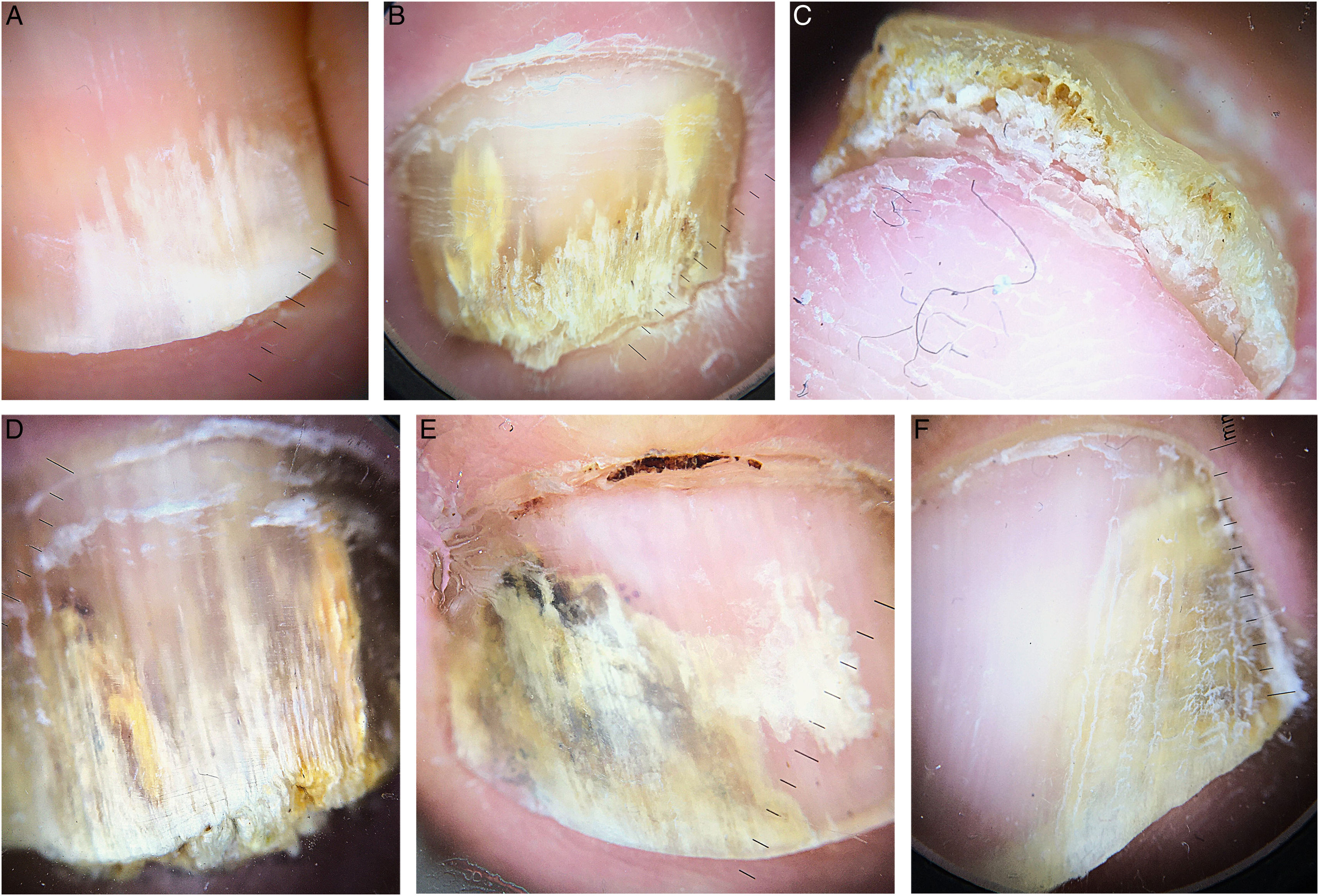
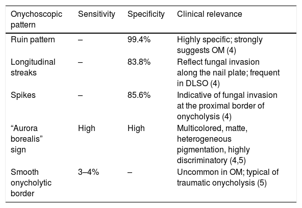
Nail dermoscopy allows identification of characteristic morphological patterns that help differentiate OM from other nail dystrophies. Since the initial descriptions by Piraccini et al., signs such as a jagged proximal edge with spikes and yellowish or brown longitudinal streaks have become established as consistent diagnostic criteria, with sensitivity rates of 77–86% and specificity rates of 95–96%31 (Fig. 4). In DLSO, the most frequent findings include the ruin pattern, longitudinal streaks, spikes at the proximal margin of onycholysis, subungual hyperkeratosis, and an irregular distal termination (Table 1). In fungal melanonychia, matte multicolor pigmentation, homogeneous nonlongitudinal pigmentation, and white or yellow streaks predominate, together with superficial transverse striation, scales, and distal linear and reverse triangular patterns related to distal fungal progression.32–35 These findings allow differential diagnosis from melanocytic melanonychia, reducing unnecessary invasive procedures.

Onychoscopic patterns in onychomycosis.
| Onychoscopic pattern | Sensitivity | Specificity | Clinical relevance |
|---|---|---|---|
| Ruin pattern | – | 99.4% | Highly specific; strongly suggests OM (4) |
| Longitudinal streaks | – | 83.8% | Reflect fungal invasion along the nail plate; frequent in DLSO (4) |
| Spikes | – | 85.6% | Indicative of fungal invasion at the proximal border of onycholysis (4) |
| “Aurora borealis” sign | High | High | Multicolored, matte, heterogeneous pigmentation, highly discriminatory (4,5) |
| Smooth onycholytic border | 3–4% | – | Uncommon in OM; typical of traumatic onycholysis (5) |
Although KOH microscopy (10–30%) and fungal culture remain reference techniques, they have limitations. KOH is rapid and inexpensive but does not identify fungal species or viability, with a sensitivity rate of 61% and specificity rate of 95%.36,37 Culture confirms viability and identifies the pathogen but has a low sensitivity rate (30–57%) and delayed results, with a specificity rate of 99%.36 Both methods are affected by previous treatments, insufficient samples, and contamination, which has prompted the use of complementary techniques such as histology and PCR.31,34
Nail histopathology using clippings stained with PAS or Grocott-Gomori is sensitive and rapid and allows fungal invasion to be differentiated from colonization. A sample≥4mm is recommended to optimize diagnosis.38 It has sensitivity rates of 85.9–88.9% and specificity rates of 82.4–100%, with results available within a few days.36,39 Hyphal morphology may suggest the type of pathogen, although it does not identify species or viability.40 It is essential for confirming infection and ruling out other nail dystrophies.
Real-time PCR allows identification of dermatophytes, NDMs, and Candida spp. within 24–48h, with a sensitivity rate of 88.5% and a specificity rate of 100%, detecting mixed infections even in the presence of contaminants.36 Its limitations include cost, the need for specialized personnel, and the inability to distinguish viable from residual DNA, which limits its use in immediate follow-up. Nevertheless, it outperforms culture in detecting mixed infections and NDMs.41
The rapid antigen test based on immunochromatography allows immediate diagnosis of tinea unguium, with a sensitivity rate of 97.2% and specificity rate of 80.7% compared with culture, even in samples with a low fungal burden.42 Its low cost and ease of use make it useful as a screening test, although it does not detect yeasts or NDMs.
AI applied to OM diagnosis has shown accuracies greater than 90%, sensitivity rates of 75–96%, and specificity rate of 92–98% in clinical and dermoscopic images.43–45 It improves the performance of digitized KOH (up to 88.8%) and PAS histopathology (sensitivity rates of 94%, specificity rates of 98%).44 However, it depends on image quality and fungal burden, does not identify species, and does not integrate relevant clinical variables, meaning that culture remains necessary.44–46
Other emerging techniques, such as flow cytometry, MALDI-TOF mass spectrometry, and confocal microscopy, provide rapid and accurate diagnoses in specific settings, but their routine use remains limited by cost, availability, and lack of standardization.6,47
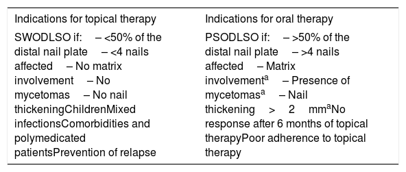
TreatmentTherapeutic management of OM needs to be optimized; it is essential to avoid empirical treatments, ensure clinical and mycological cure, and prevent recurrences.48 Treatment choice should be individualized according to clinical characteristics, infection severity, and personal factors such as age or comorbidities49 (Table 2). The main pharmacological strategies include systemic, topical, and physical treatment.
Treatment indications.
| Indications for topical therapy | Indications for oral therapy |
|---|---|
| SWODLSO if:– <50% of the distal nail plate– <4 nails affected– No matrix involvement– No mycetomas– No nail thickeningChildrenMixed infectionsComorbidities and polymedicated patientsPrevention of relapse | PSODLSO if:– >50% of the distal nail plate– >4 nails affected– Matrix involvementa– Presence of mycetomasa– Nail thickening>2mmaNo response after 6 months of topical therapyPoor adherence to topical therapy |
Oral antifungal agents have demonstrated significantly higher cure rates with shorter treatment times than topical treatments, but their use may be limited in some patients.4,50
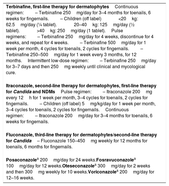
Terbinafine is the first-line drug for dermatophyte OM, with better cure rates than azoles and fewer recurrences.51,52 The usual regimen is continuous administration of 250mg daily and, although it appears superior, other studies have found comparable efficacy with pulse regimens, with lower cost and low toxicity.53–56 Intermittent low-dose regimens have been proposed, particularly for older or polymedicated patients, with good empirical results57 (Table 3).
Systemic therapy.
| Terbinafine, first-line therapy for dermatophytesContinuous regimen:– Terbinafine 250mg/day for 3–4 months for toenails, 6 weeks for fingernails.– Children (off label)<20kg: 62.5mg/day (¼ tablet).20–40kg: 125mg/day (½ tablet).>40kg: 250mg/day (1 tablet).Pulse regimens:– Terbinafine 250mg/day for 4 weeks, discontinue for 4 weeks, and repeat for 4 weeks.– Terbinafine 500mg/day for 1 week per month, 4 cycles for toenails, 2 cycles for fingernails.– Terbinafine 250–500mg/day for 1 week every 3 months, for 12 months.Intermittent low-dose regimen:– Terbinafine 250mg/day for 3–7 days and then 250mg weekly until clinical and mycological cure. |
| Itraconazole, second-line therapy for dermatophytes, first-line therapy for Candida and NDMsPulse regimen:– Itraconazole 200mg every 12h for 1 week per month, 3–4 cycles for toenails, 2 cycles for fingernails.– Children (off label) 5mg/kg/day for 1 week per month, 3–4 cycles for toenails, 2 cycles for fingernails.Continuous regimen:– Itraconazole 200mg/day for 3–4 months for toenails, 6 weeks for fingernails. |
| Fluconazole, third-line therapy for dermatophytes/second-line therapy for Candida– Fluconazole 150–450mg weekly for 12 months for toenails, 6 months for fingernails. |
| Posaconazolea 200mg/day for 24 weeks.Fosravuconazolea 100mg/day for 12 weeks.Oteseconazolea 300mg/day for 2 weeks and then 300mg weekly for 10 weeks.Voriconazolea 200mg/day for 12–16 weeks. |
Although itraconazole is considered second-line therapy for dermatophyte OM, it is the drug of choice for infections caused by Candida or NDMs. It is usually prescribed in pulses of 200mg every 12h for one week per month, although continuous treatment with 200mg daily provides superior results50 (Table 3).
Fluconazole is a third-line alternative for dermatophytes and second-line therapy after itraconazole for Candida infections, with weekly doses of 150–450mg, with better results in fingernails and at the higher doses.1,18 (Table 3).
Griseofulvin has fallen out of use because of lower efficacy and greater toxicity.18
Among the newer antifungals, fosravuconazole has the most evidence in OM; it is superior to topical treatments and has an adequate safety profile.58 Posaconazole, oteseconazole, or voriconazole, with limited evidence and no standardized regimens, are reserved for cases with absolute contraindication to or repeated failure of conventional antifungal agents59 (Table 3).
Acitretin combined with itraconazole improves outcomes but with a higher risk of hepatotoxicity.60
Although the most common adverse effects of antifungal agents are usually GI disturbances and headache, hepatotoxicity and drug interactions are the main limitations. Terbinafine has fewer drug interactions than itraconazole but a greater risk of hepatotoxicity. Recent studies show low rates of reversible transaminase elevation, and routine laboratory monitoring would not be necessary in patients without a history of liver disease, although it is recommended in those older than 65 years or on multiple drugs.56,61–63 Itraconazole should be avoided in patients with heart disease and may cause serious drug interactions, increasing benzodiazepine and statin concentrations with a risk of rhabdomyolysis, and should be avoided in patients with risk factors for QT prolongation.64–66
Topical therapyAlthough topical therapies have an excellent safety profile, they have penetration limitations and require treatment for 12–18 months. As monotherapy, they are indicated in mild forms. The nail lacquer formulation allows better diffusion and easier application (Table 2).
Amorolfine 5%, applied 1–2 times/week, and ciclopirox 8%, applied daily, have similar efficacy, although 8% ciclopirox hydrolacquers with hydroxypropyl chitosan provide better complete cure rates (clinical and mycological).50,67
Efinaconazole 10%, available only in some countries, shows results similar to oral itraconazole, high penetration even in dermatophytomas, and a broad spectrum of activity with greater efficacy than other topical agents.68–70
Penetration of topical therapies can be improved by combining chemical penetration enhancers such as urea, salicylic acid, or hydroxypropyl chitosan, or by using physical procedures such as iontophoresis, nail plate microperforations, and, in the future, incorporation of nanotechnology.
DebridementRemoval of as much diseased nail as possible improves the likelihood of cure, always in combination with topical or oral treatment.71 In dermatophytomas, it is essential and, when combined with terbinafine, appears to be the best option.69 It is also mandatory in NDM infections, in which the combination of debridement and topical antifungals for prolonged periods is the preferred strategy.71 Debridement may be performed chemically using keratolytics such as 40% urea, mechanically by trimming or burring the nail, or surgically by partial nail avulsion, avoiding total avulsion to minimize the possibility of dystrophy.6
Devices in the treatment of onychomycosisNumerous studies have been published on devices aimed at inhibiting fungal growth or facilitating the permeability of antifungal agents, but with low methodological quality and modest results.72,73
Although lasers have demonstrated clinical improvements, there is no evidence on their sustainability or mycological cure.74 Although 93% of patients treated with monotherapy do not achieve mycological cure, lasers do improve the results of topical therapy, which is of interest in the management of NDM infections and in cases of resistance to or contraindication for systemic therapy75
Photodynamic therapy has a broad spectrum of activity, acts on biofilms, and appears to provide good mycological results. It is considered an adjunct to topical therapies.74
Alternative therapiesAmong the many therapies that have been promoted are thymol and essential oils, which have demonstrated antifungal, antioxidant, and anti-inflammatory activity. Tea tree oil has been widely studied but without conclusive evidence.76 Nitric acid 60% applied in the clinic or sodium hypochlorite as 1% soaks or as direct weekly application are other alternatives, although without proven efficacy.77
Combination therapyThe combination of oral therapy, topical therapy, and debridement offers the best chances of success. Combined oral and topical therapy is superior to monotherapy, prevents recurrence and resistance, increases efficacy in mixed infections, and reduces biofilms, thereby decreasing treatment duration, cost, and toxicity.48,78,79
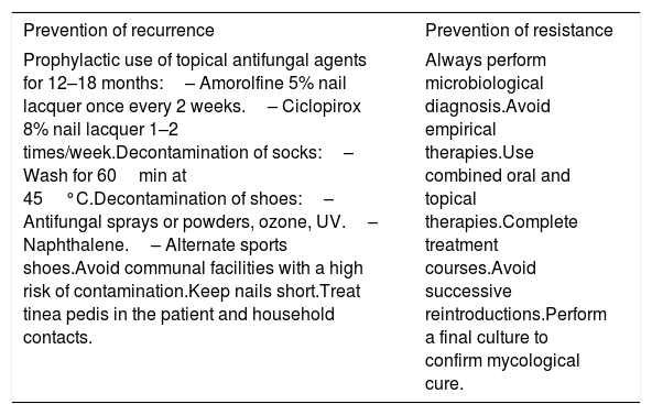
Prevention of recurrenceRecurrences are less frequent in patients treated with terbinafine or combination therapy.4,6 Patients with diabetes, immunosuppression, severe clinical forms, or tinea pedis have higher recurrence rates. Prevention may be achieved with topical antifungal agents accompanied by hygienic measures80,78,81,82 (Table 4).
Measures for the prevention of recurrence and resistance.
| Prevention of recurrence | Prevention of resistance |
|---|---|
| Prophylactic use of topical antifungal agents for 12–18 months:– Amorolfine 5% nail lacquer once every 2 weeks.– Ciclopirox 8% nail lacquer 1–2 times/week.Decontamination of socks:– Wash for 60min at 45°C.Decontamination of shoes:– Antifungal sprays or powders, ozone, UV.– Naphthalene.– Alternate sports shoes.Avoid communal facilities with a high risk of contamination.Keep nails short.Treat tinea pedis in the patient and household contacts. | Always perform microbiological diagnosis.Avoid empirical therapies.Use combined oral and topical therapies.Complete treatment courses.Avoid successive reintroductions.Perform a final culture to confirm mycological cure. |
Lack of response reflects clinical or microbiological failure. Clinical failure may be due to diagnostic error, inadequate treatment, extensive disease, or immunosuppression. The combination of terbinafine and itraconazole does not appear to improve outcomes.80,83 NDM and mixed infections respond less well and, although itraconazole appears to be the best oral agent, topical treatments combined with debridement are more effective.84 In severe cases or those with limited response, booster therapy with an additional pulse at 6 or 9 months after treatment has been proposed, but its efficacy is not well established.80,85 In chronic onychomycosis, repeated courses of treatment may be necessary, without overlooking the possibility of microbiological resistance.
Terbinafine resistance is a growing and probably underestimated concern; most cases are associated with mutations in the squalene epoxidase (SQLE) gene.86 Antifungal susceptibility testing for dermatophytes is not routinely performed and has interpretative limitations; detection of mutations in the SQLE gene (F397L, L393F, and A448T) may be more informative, although access is limited.87,88 There are no consensus guidelines for management; the strategy should focus on prevention and, when resistance is suspected and clinical failure has been ruled out, susceptibility testing should be performed and treatment changed to itraconazole and/or topical treatments for which resistance has not been demonstrated.89 (Table 4). As a last option, newer antifungals could be considered and, although evidence in resistance is limited, cases associated with reduced susceptibility to itraconazole have been observed and must be taken into consideration.90
Special situationsIn patients with diabetes, improving glycemic control is important and terbinafine is preferred because of the lower risk of hypoglycemia. In patients with renal or hepatic disease, topical treatments are preferable; terbinafine would require dose adjustment in patients with nephropathy.66,91 In immunocompromised patients, systemic therapy is recommended, preferably terbinafine, with caution if CD4 counts are <500, and debridement should be avoided.92 Terbinafine may worsen psoriasis, and itraconazole is recommended in these cases.93 Oral treatment in older patients will be terbinafine because it has fewer serious interactions, combined with topical agents.
In childhood, topical agents are preferred because of their safety, although studies report low cure rates. Ciclopirox 8% nail lacquers are approved for patients older than 12 years. There is off-label experience with terbinafine and itraconazole, with good cure rates in small samples, but terbinafine is preferred with laboratory monitoring given the limited experience in these patients13 (Table 4). During pregnancy and breastfeeding, ciclopirox 8% nail lacquer combined with mechanical debridement is recommended, and during breastfeeding the addition of laser therapy may be considered.91
Ethical considerationsThe present work was conducted in full compliance with the ethical principles established in the Declaration of Helsinki. Given the nature of the study, approval by a research ethics committee was not required.
Informed consentOral informed consent was obtained from the patient for the acquisition and publication of the clinical images included in this article, with anonymity guaranteed at all times.
Use of generative artificial intelligence in scientific writingGenerative artificial intelligence was used as linguistic support in drafting the manuscript. The authors assume full responsibility for the scientific content.
FundingThis work was funded by Almirall, which provided financial support for preparation of the manuscript.
Conflicts of interestL. Navarro Campoamor has served as a medical advisor and/or speaker for Almirall and Viñas.
M. Iglesias-Sancho has served as a medical advisor and/or speaker and/or has received research funding from Almirall, AbbVie, UCB, Janssen, ISDIN, and Novartis.
E. Jiménez Blázquez has served as a medical advisor and/or speaker for Almirall and Viñas.
Uncited reference15.