


Leprosy, also known as Hansen disease, is an infectious disease caused by Mycobacterium leprae and Mycobacterium lepromatosis, with manifestations involving the skin and peripheral nerves, among other organs. Assessing the prevalence and incidence of leprosy is challenging because of its low occurrence and the possibility of delayed diagnosis. Most registered and newly detected cases originate in endemic regions of Southeast Asia and South America.1,2 However, increasing population mobility means that cases may be diagnosed anywhere.3–5
We report two cases of leprosy recently diagnosed in Barcelona, Spain.
Case 1A 24-year-old woman originally from Bolivia who had been living in Spain for 2 years presented to our department with skin lesions of 14 months’ duration. The lesions initially appeared on the trunk and subsequently became generalized. She denied unprotected sexual intercourse and reported no travel during the preceding 2 years. Previous treatment with antifungal agents and topical corticosteroids had resulted in no improvement.
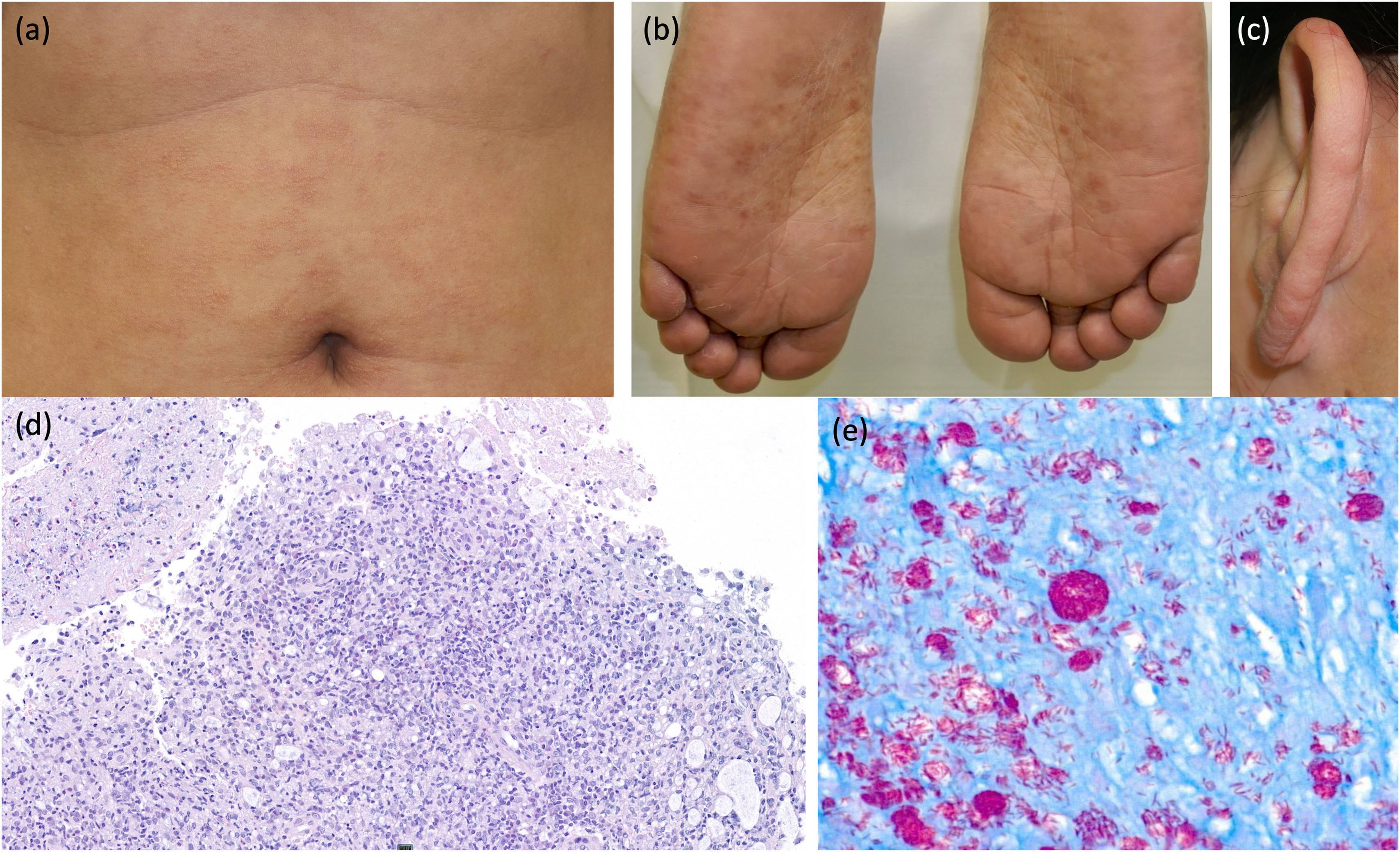
Physical examination revealed millimetric papules distributed across the trunk, papules and nodules on the extremities, face, and ears, and hyperpigmented papules on the soles (Fig. 1). Neurological examination revealed hypoesthesia in a stocking distribution affecting both lower extremities, without palpable nerve thickening.
Case 1. Millimetric erythematous papules distributed in groups across the abdomen, with intervening areas of unaffected skin (a). Hyperpigmented papules involving the soles (b). Firm nodules involving the left auricle (c). Histopathological examination of a nodule on the lower extremity revealed a diffuse histiocytic infiltrate with a poorly defined granulomatous appearance, predominantly involving the mid and deep dermis, with plasma cells and occasional eosinophils (hematoxylin–eosin [H&E] stain, ×400) (d). Fite–Faraco staining revealed numerous bacilli, both scattered and clustered, within the cytoplasmic vacuoles of histiocytes (globi) (Fite–Faraco stain, ×400) (e).
Laboratory testing revealed elevated inflammatory markers. Serological tests for HIV, hepatotropic viruses, and syphilis were negative. Tuberculosis was ruled out on the basis of a negative interferon-gamma release assay (IGRA).
Histopathological examination of a nodule on the lower extremity revealed a poorly defined granulomatous infiltrate and histiocytes containing cytoplasmic vacuoles. Fite–Faraco staining demonstrated numerous bacilli (Fig. 1).
A diagnosis of multibacillary (MB) leprosy was established. Multidrug therapy (MDT) consisting of rifampicin, dapsone, and clofazimine was prescribed for 12 months.
Case 2A 48-year-old woman who had been living with the patient described in Case 1 for the preceding 18 months presented to our department for evaluation of possible leprosy-associated skin lesions.
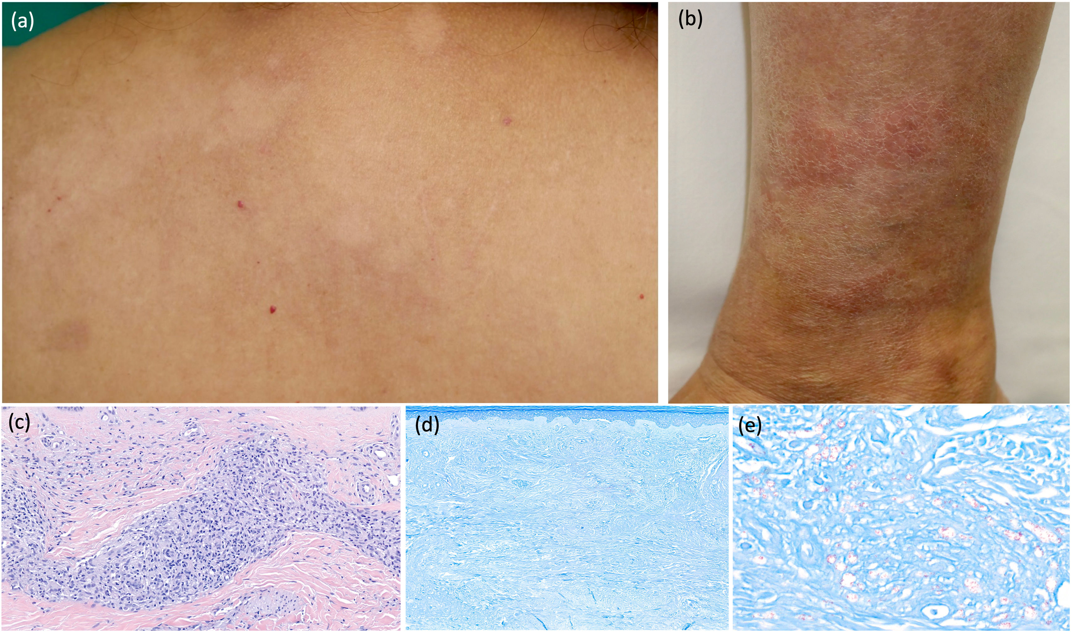
A complete physical examination revealed an erythematous plaque with raised borders and a hypopigmented, anesthetic center on the left lower extremity. Hypopigmented macules resembling pityriasis versicolor, with preserved sensation, were also observed on the back (Fig. 2).
Case 2. Erythematous plaque with a raised border and a hypopigmented center on the left lower extremity (a). Hypopigmented macules with indistinct borders resembling pityriasis versicolor, involving the upper back (b). Histopathological examination of the active border of the plaque on the leg revealed nonnecrotizing granulomatous dermatitis surrounding nerves and blood vessels, composed of epithelioid histiocytes, lymphocytes, and plasma cells (H&E stain, ×400) (c). Ziehl–Neelsen staining revealed no bacilli (×200) (d). In contrast, Fite–Faraco staining demonstrated numerous scattered bacilli in the dermis (×400) (e).
Laboratory tests and serological studies revealed no abnormalities. The IGRA result was negative.
Histopathological examination of the active border of the plaque on the leg revealed nonnecrotizing granulomatous dermatitis surrounding peripheral nerves. Gram, Ziehl–Neelsen, and Grocott staining revealed no microorganisms. However, Fite–Faraco staining, performed at our request, demonstrated numerous bacilli (Fig. 2).
These findings confirmed the diagnosis of MB leprosy. The patient was started on the same MDT regimen prescribed for the first patient.
In the current era of globalization, leprosy is increasingly being diagnosed in regions where no autochthonous cases have been reported in recent years. This is illustrated by our findings and recent reports of leprosy in locations such as Florida.4 Although leprosy is believed to be transmitted through respiratory droplets from untreated patients with lepromatous leprosy,1 the risk of acquiring the disease following close contact has been estimated at approximately 25%.6 Nevertheless, most exposed individuals do not develop the disease because of multiple factors, including immune status and genetic susceptibility. The incubation period varies considerably, ranging from 2 years to more than a decade.2
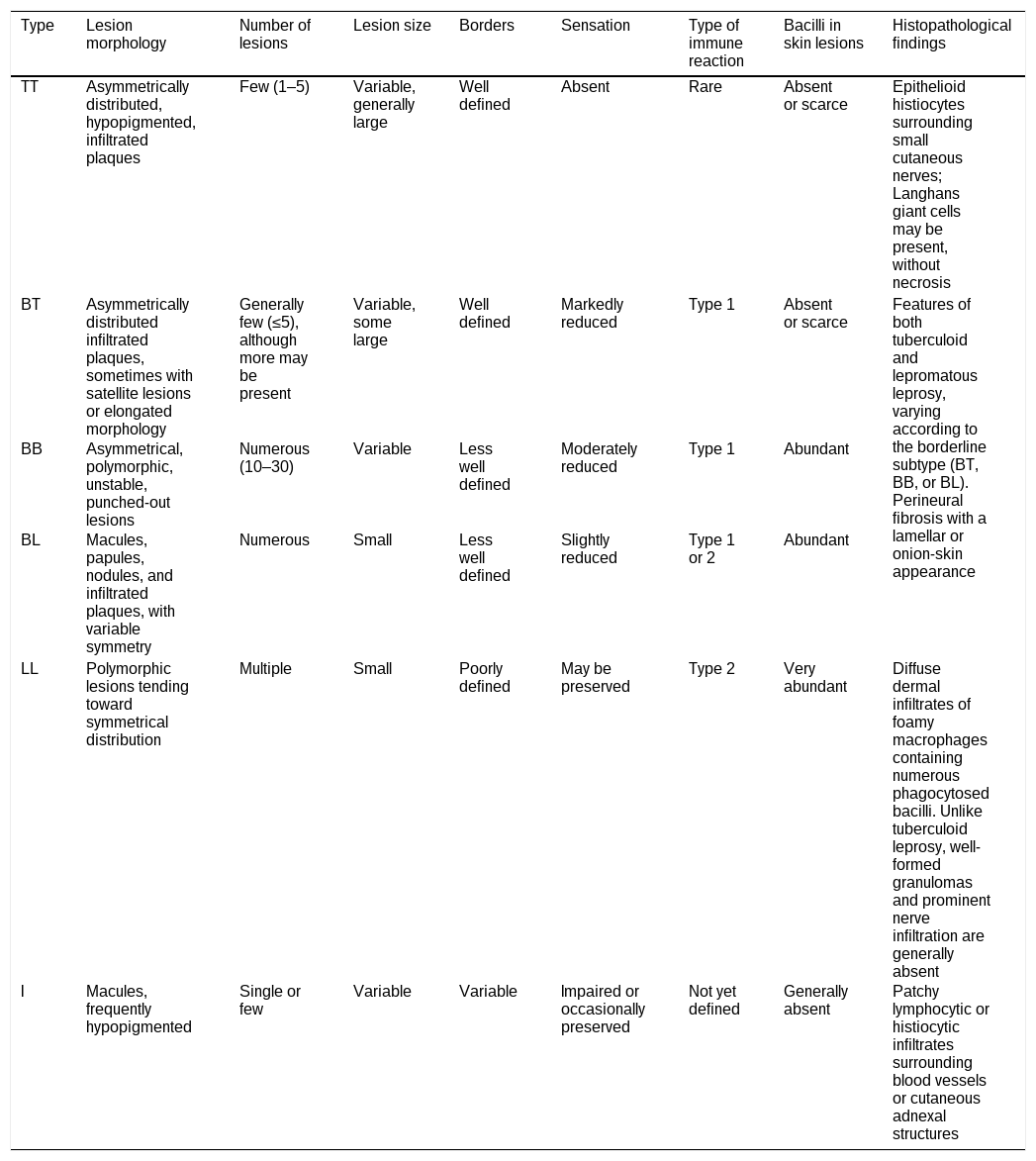
Leprosy encompasses a spectrum of clinical, immunological, and histopathological signs determined by individual susceptibility to the causative pathogens.1 The Ridley–Jopling classification reflects this complexity and correlates with bacillary burden7 (Table 1).
Classification of leprosy according to the Ridley–Jopling system across the disease spectrum.
| Type | Lesion morphology | Number of lesions | Lesion size | Borders | Sensation | Type of immune reaction | Bacilli in skin lesions | Histopathological findings |
|---|---|---|---|---|---|---|---|---|
| TT | Asymmetrically distributed, hypopigmented, infiltrated plaques | Few (1–5) | Variable, generally large | Well defined | Absent | Rare | Absent or scarce | Epithelioid histiocytes surrounding small cutaneous nerves; Langhans giant cells may be present, without necrosis |
| BT | Asymmetrically distributed infiltrated plaques, sometimes with satellite lesions or elongated morphology | Generally few (≤5), although more may be present | Variable, some large | Well defined | Markedly reduced | Type 1 | Absent or scarce | Features of both tuberculoid and lepromatous leprosy, varying according to the borderline subtype (BT, BB, or BL). Perineural fibrosis with a lamellar or onion-skin appearance |
| BB | Asymmetrical, polymorphic, unstable, punched-out lesions | Numerous (10–30) | Variable | Less well defined | Moderately reduced | Type 1 | Abundant | |
| BL | Macules, papules, nodules, and infiltrated plaques, with variable symmetry | Numerous | Small | Less well defined | Slightly reduced | Type 1 or 2 | Abundant | |
| LL | Polymorphic lesions tending toward symmetrical distribution | Multiple | Small | Poorly defined | May be preserved | Type 2 | Very abundant | Diffuse dermal infiltrates of foamy macrophages containing numerous phagocytosed bacilli. Unlike tuberculoid leprosy, well-formed granulomas and prominent nerve infiltration are generally absent |
| I | Macules, frequently hypopigmented | Single or few | Variable | Variable | Impaired or occasionally preserved | Not yet defined | Generally absent | Patchy lymphocytic or histiocytic infiltrates surrounding blood vessels or cutaneous adnexal structures |
TT: polar tuberculoid; BT: borderline tuberculoid; BB: mid-borderline; BL: borderline lepromatous; LL: polar lepromatous; I: indeterminate.
Adapted from:
1. Ramos-e-Silva M, Ribeiro de Castro MC. Mycobacterial infections. In: Bolognia JL, Schaffer JV, Cerroni L, eds. Dermatology. 4th ed. Elsevier; 2018. p. 1296–303.
2. Ridley DS, Jopling WH. Classification of leprosy according to immunity. A five-group system. Int J Lepr Other Mycobact Dis. 1966;34(3):255–73.
3. Hamodat M. Leprosy. Pathology Outlines. https://www.pathologyoutlines.com/topic/skinnontumorleprosy.html Accessed 10.9.23.
For practical purposes, the World Health Organization recommends classifying leprosy according to the number of skin lesions when making treatment decisions in endemic regions (paucibacillary [PB] leprosy if ≤5 lesions).8 However, this approach may lead to misclassification, as demonstrated in published studies.7,9 Histopathological examination is therefore essential when available.
The choice of staining technique is also important. Fite–Faraco staining is more sensitive than Ziehl–Neelsen staining for diagnosing leprosy,10 as illustrated by our findings in Case 2.
Our cases, diagnosed in a nonendemic region, highlight the importance of including leprosy in the differential diagnosis of dyschromic, annular, nodular, or infiltrative skin lesions, particularly when they do not respond to treatment. Assessment of cutaneous sensation and Fite–Faraco staining of biopsy specimens are essential diagnostic tools. Early diagnosis and treatment not only prevent disability but also contribute to public health protection.
FundingThis work received no specific funding.
Informed consentWritten informed consent was obtained from both patients for the publication of their clinical information and images.
Conflict of interestThe authors declare that they have no conflict of interest.