


Mogamulizumab is a monoclonal antibody that specifically targets CCR4 and has been approved for patients with mycosis fungoides (MF) and Sézary syndrome (SS) who have failed at least 1 prior systemic therapy. This approval was based on the findings of the MAVORIC trial, which highlighted several adverse effects associated with disruption of immune regulation, particularly involving CD4+ T cells. Among these, drug-induced skin reactions were reported in 24% of cases.1 However, more recent studies suggest that these cutaneous adverse events may be more prevalent than initially recognized, with some reports indicating an incidence of up to 63%. Notably, growing evidence suggests that these dermatologic reactions may correlate with a more favorable prognosis and enhanced therapeutic response.2,3 One of the main challenges associated with these cutaneous manifestations—collectively referred to as mogamulizumab-associated rash (MAR)—is their clinical resemblance to MF/SS lesions, which complicates accurate diagnosis.4
At our tertiary referral center, Hospital Universitario 12 de Octubre (Madrid, Spain), 53 patients (n=53) with SS or MF were undergoing treatment with mogamulizumab. Among them, 20 patients (n=20) developed MAR with different clinical patterns. A remarkable feature was the novel presentation of localized, erythematous, scaly plaques on the upper backobserved in 6 patients (n=6). A skin biopsy was performed in all patients to rule out lymphoma involvement and confirm the diagnosis of a mogamulizumab-induced drug reaction. The clinical and histopathologic features were described and compared with the existing literature.
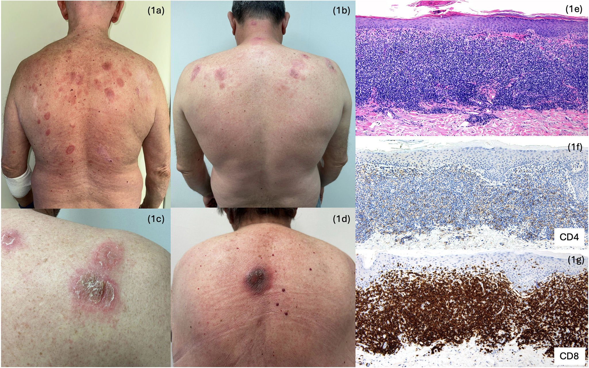
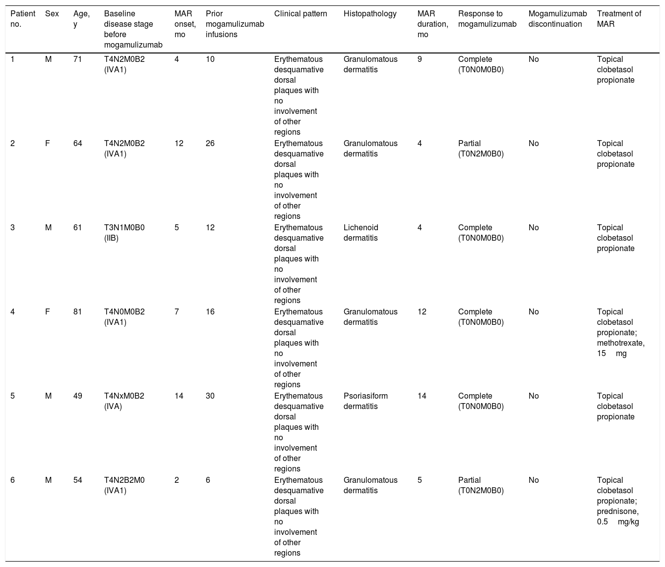
The series included a total of 6 patients (4 men and 2 women) with diagnoses of mycosis fungoides (n=4) and Sézary syndrome (n=2). The median age was 65 years (range, 49–81 years). Five patients presented with stage IV disease at the initiation of mogamulizumab; the remaining patient had stage IIB disease. The median time to onset of the dorsal plaques was 7 months (range, 1–14 months), with a median of 17 mogamulizumab infusions administered before rash development (range, 6–30 infusions). At the time of lesion onset, all patients were in complete or partial response in the skin, lymph nodes, and peripheral blood. Clinically, the rash appeared as infiltrated, scaly, erythematous plaques located on the upper back (Fig. 1A–D). No skin lesions were detected beyond the dorsal region. The most characteristic histopathologic finding, present in 4 patients (n=4), was a granulomatous inflammatory infiltrate (Fig. 1E), with inversion of the CD4/CD8 ratio on immunohistochemistry (IHC), showing a marked predominance of CD8+ lymphocytes and abundant eosinophils (Fig. 1F, G). Other histopathologic patterns were lichenoid (n=1) and psoriasiform (n=1). Management of these newly developed dorsal lesions included topical clobetasol propionate (n=6), prednisone 0.5mg/kg (n=1), and methotrexate 15mg (n=1). Mogamulizumab treatment was continued in all patients, and at the last follow-up, 4 individuals were in complete remission of their underlying disease and 2 were in partial response (Table 1).

Clinical characteristics of patients with MF who developed dorsal plaques following treatment with mogamulizumab.
| Patient no. | Sex | Age, y | Baseline disease stage before mogamulizumab | MAR onset, mo | Prior mogamulizumab infusions | Clinical pattern | Histopathology | MAR duration, mo | Response to mogamulizumab | Mogamulizumab discontinuation | Treatment of MAR |
|---|---|---|---|---|---|---|---|---|---|---|---|
| 1 | M | 71 | T4N2M0B2 (IVA1) | 4 | 10 | Erythematous desquamative dorsal plaques with no involvement of other regions | Granulomatous dermatitis | 9 | Complete (T0N0M0B0) | No | Topical clobetasol propionate |
| 2 | F | 64 | T4N2M0B2 (IVA1) | 12 | 26 | Erythematous desquamative dorsal plaques with no involvement of other regions | Granulomatous dermatitis | 4 | Partial (T0N2M0B0) | No | Topical clobetasol propionate |
| 3 | M | 61 | T3N1M0B0 (IIB) | 5 | 12 | Erythematous desquamative dorsal plaques with no involvement of other regions | Lichenoid dermatitis | 4 | Complete (T0N0M0B0) | No | Topical clobetasol propionate |
| 4 | F | 81 | T4N0M0B2 (IVA1) | 7 | 16 | Erythematous desquamative dorsal plaques with no involvement of other regions | Granulomatous dermatitis | 12 | Complete (T0N0M0B0) | No | Topical clobetasol propionate; methotrexate, 15mg |
| 5 | M | 49 | T4NxM0B2 (IVA) | 14 | 30 | Erythematous desquamative dorsal plaques with no involvement of other regions | Psoriasiform dermatitis | 14 | Complete (T0N0M0B0) | No | Topical clobetasol propionate |
| 6 | M | 54 | T4N2B2M0 (IVA1) | 2 | 6 | Erythematous desquamative dorsal plaques with no involvement of other regions | Granulomatous dermatitis | 5 | Partial (T0N2M0B0) | No | Topical clobetasol propionate; prednisone, 0.5mg/kg |
The proposed pathophysiologic mechanism underlying MAR involves loss of regulation of CD8+ T cells by regulatory T cells, which also express CCR4. This dysregulation results in increased CD8+ T-cell activation.5 MAR is a relatively common cutaneous adverse event in patients treated with mogamulizumab and can present with variable clinical features. Prompt recognition and histopathologic evaluation of newly developed skin lesions are essential for accurate diagnosis and effective clinical management. In this context, we describe a clinical variant of MAR that may serve as a valuable diagnostic clue. Treatment of MAR should be guided by its severity; in most cases, continuation of mogamulizumab alongside appropriate adjunctive therapy is recommended, particularly given the association of MAR with favorable treatment outcomes.
Funding sourcesNone.
Conflicts of interestNone declared.