

A 65-year-old woman with no drug allergies and a medical history of type II diabetes mellitus, dyslipidemia, and HLA-B27-positive seronegative spondyloarthropathy, was referred from the hematology department for progressive asymptomatic thickening of the lip that had begun several months earlier. The patient was undergoing tests for moderate iron deficiency anemia and a monoclonal immunoglobulin (Ig) G λ band. Prior tests had revealed no other findings of interest.
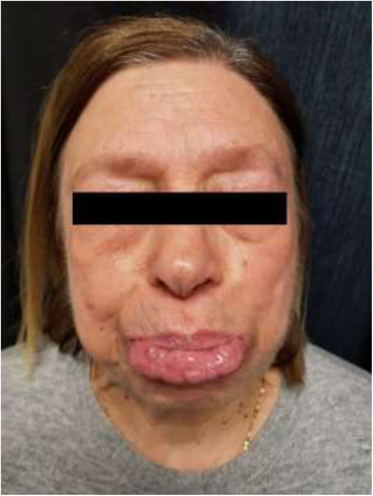
Physical ExaminationThe patient’s upper lip was thickened and hard to the touch, without associated ulcers (Fig. 1). Neither locoregional lymphadenopathy nor hepatosplenomegaly were palpable. Examination of the skin and mucosa revealed no other lesions.
Histopathology
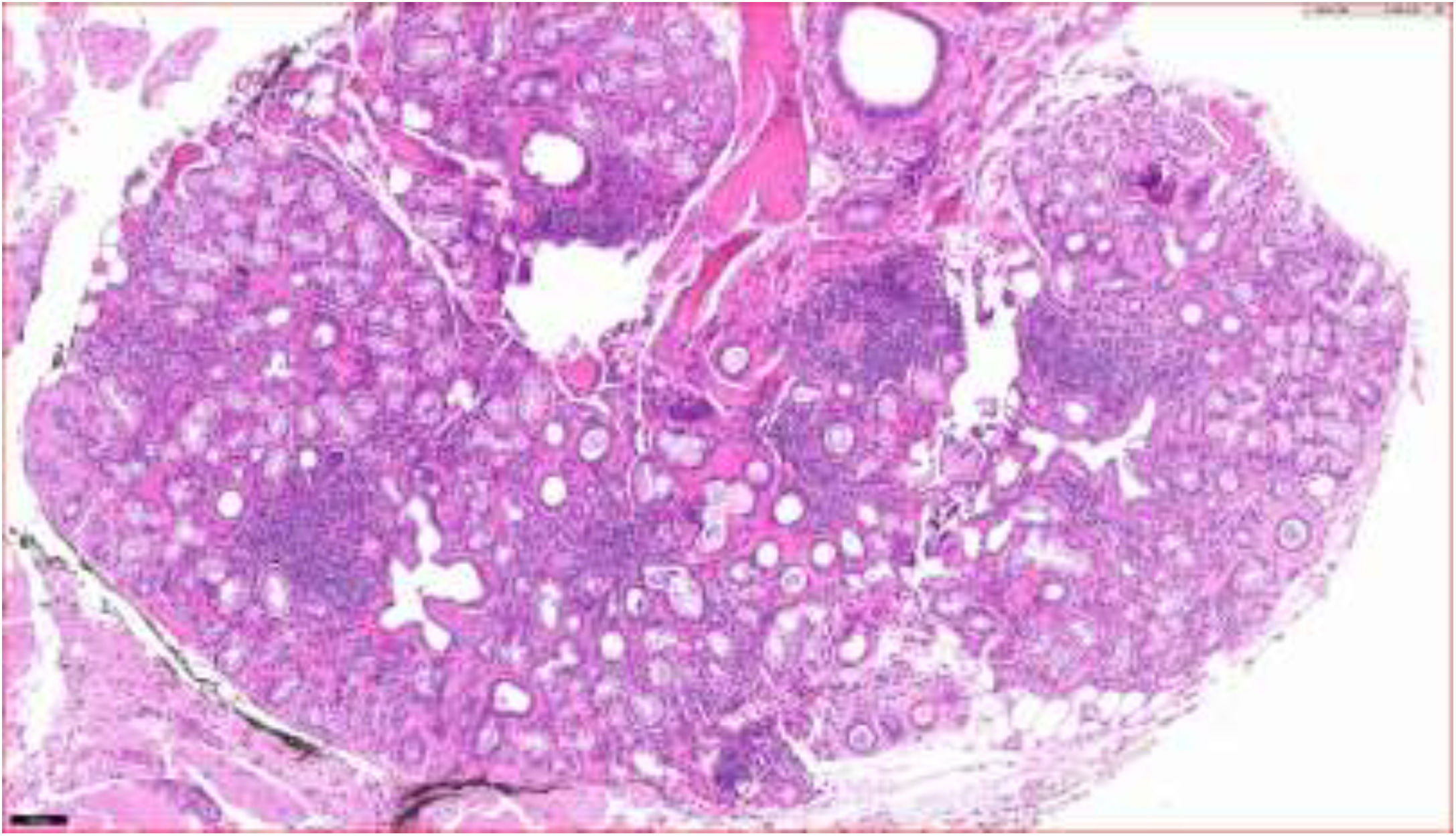
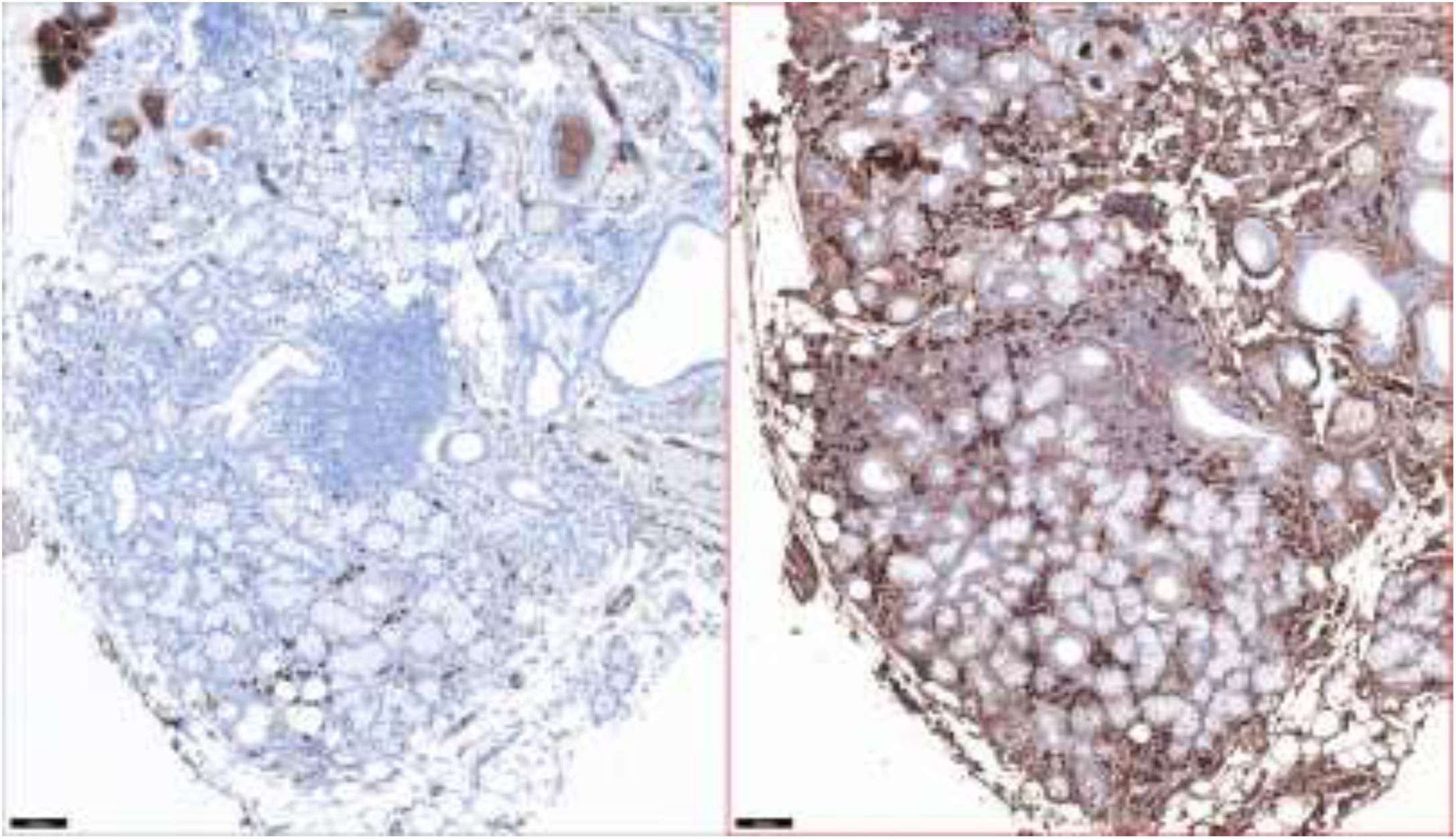
Histopathology showed lymphoid clusters consisting mainly of plasma cells with a periglandular distribution (Fig. 2). Immunohistochemistry was positive for CD20 and negative for IgG and IgG4, and revealed monoclonal λ light chains but was negative for κ light chains (Fig. 3).
Additional Tests
 G κ light chain. Right, positive staining for IgG λ light chain.")
Additional tests revealed a normal complete blood count and a normal biochemical profile. Quantification of IgG λ light chain in serum continued to show elevated levels (3.20 g/dL). The results of the autoimmunity study were normal. The results of a bone marrow aspiration (BMA) biopsy were within the normal range. The first positron emission tomography–computed tomography (PET–CT) scan showed foci of mild-to-moderate metabolic activity associated with paratracheal lymphadenopathy suggestive of benign inflammatory disease.
What Is Your Diagnosis?DiagnosisLymphocytic hyperplasia with monoclonal IgG λ plasmacytosis.
Clinical Course and TreatmentBMA biopsy was repeated 3 months later and revealed an increase in plasma cells (2.5%) and clonal B lymphocytes with weak cytoplasmic expression of IgG λ light chain. A second PET–CT scan was performed 6 months later and compared with previous images. The scan showed findings compatible with adenopathies suggestive of lymphomatous spread and a probable diagnosis of IgG λ lymphoplasmacytic lymphoma. Treatment with cycles of rituximab, cyclophosphamide, and dexamethasone (RCD) resulted in a progressive decrease in the monoclonal component, but had no effect on the patient’s lip condition.
CommentMonoclonal plasmacytosis in minor salivary gland biopsies is observed in autoimmune diseases such as Sjögren syndrome, in the early stages of mucosa-associated lymphoid tissue lymphoma, and even in monoclonal gammopathy of uncertain significance.1 Lip and minor salivary gland involvement, as observed in the present case, may be the first manifestation of lymphoma with systemic compromise. Transition between lymphocytic infiltrate with apparently benign characteristics and lymphoma occurs relatively frequently, in some cases separated by intermediate stages that are difficult to classify. Therefore, clonal proliferations in clinically accessible locations (e.g. the lip, which in our patient became progressively thickened) should be evaluated with caution. It is necessary to clinically rule out processes such as granulomatous cheilitis in Melkersson-Roshental syndrome2 and Sjögren syndrome, in which these clonal proliferations have also been described, as well as IgG4-related disease,3 which was ruled out in our patient.
Histologically, features that suggest benignity include preserved acinar architecture and the presence of small lymphocytes and plasma cells in the interfollicular regions with a nondiffuse pattern distinct from that of lymphoproliferative infiltrate.4
Ultimately, our patient was diagnosed with IgG λ lymphoplasmacytic lymphoma. In 20085 the World Health Organization defined this condition as a B-cell neoplasm consisting of coexisting clonal populations of small B cells, lymphoplasmacytes, and plasma cells. It is frequently associated with a monoclonal IgM component (Waldenstrom macroglobulinemia) and less than 5% of patients present a monoclonal IgG band,6 which was observed in our patient.
FundingNo funding was received for this study.
Conflicts of interestThe authors declare that they have no conflicts of interest.
Please cite this article as: Ruiz-Villaverde R, Rueda-Villafranca B, Galvez-Moreno M. Engrosamiento labial progresivo asintomático. Actas Dermosifiliogr. 2021;112:645–646.


