

An 84-year-old man with considerable actinic damage was seen for a painless tumor on the face that had appeared several years earlier had progressively increased in size. The growth of the lesion had accelerated in the months preceding the consultation.
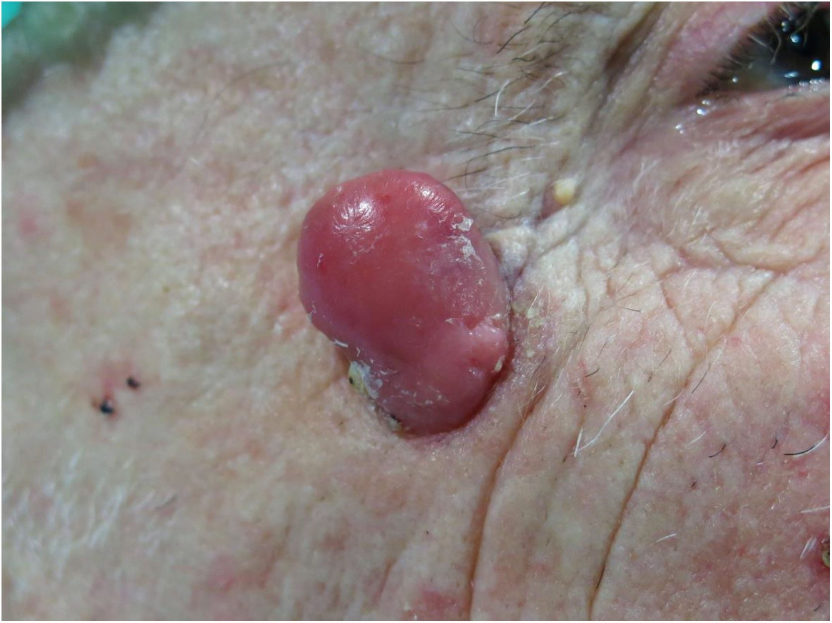
Physical ExaminationExamination of the skin revealed a pink exophytic tumor (2.5 × 2 cm) on the outer canthus of the right eye, with a smooth surface, firm consistency, and slightly infiltrated neat borders (Fig. 1).
Histopathology
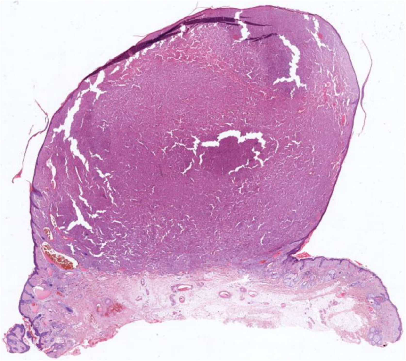
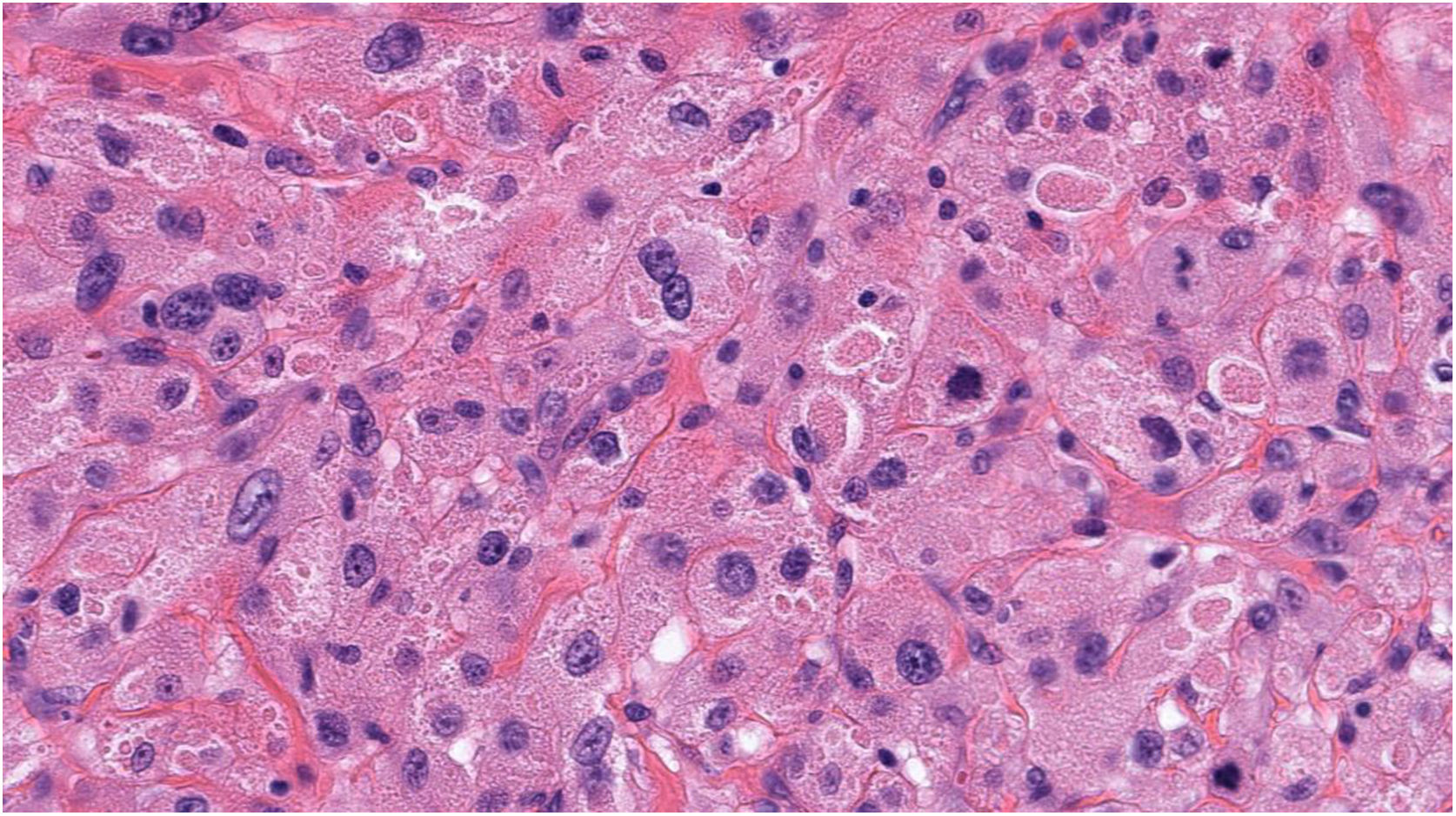
Histology revealed a well-defined dermal tumor with a polypoid appearance (Fig. 2) consisting of large cells of varied shape with abundant eosinophilic cytoplasm containing granules, nuclear pleomorphism, and some mitotic figures (Fig. 3). Immunohistochemistry showed intense reactivity to CD10 and CD68 and was negative for S-100, MelanA, HMB45, and markers of neural, epithelial, and muscular differentiation.
.")
.")
What Is Your Diagnosis?
DiagnosisPrimitive polypoid granule cell tumor.
Clinical Course and TreatmentThe tumor was fully excised and the patient has shown no signs of recurrence or metastasis after 1 year of follow-up.
CommentPrimitive polypoid granular cell tumor is a rare tumor of uncertain lineage that was first described by Le Boit et al in 1991.1 There are very few published cases in the literature, and both sexes appear to be equally affected.2 Most cases present before 50 years of age and the most common location is the trunk, followed by the extremities and the head and neck. The tumor is painless and of variable size.3
Unlike conventional granular cell tumor, it is characterized by neater borders, negative S-100 immunostaining, and more concerning cytological findings with marked nuclear pleomorphism and a higher mitotic count.4 The most problematic condition included in the differential diagnosis is granular cell atypical fibroxanthoma (FXA), which affects individuals in the same age group as our patient, has similar immunohistochemical features, and occurs in the same location and on photodamaged skin.5 However, this condition can be ruled out based on the absence of a spindle-cell area, which is characteristic of FXA, as well as less marked cellular atypia and less mitotic activity without atypical mitoses.
Diagnosis is established by exclusion, mainly based on histology and immunohistochemistry findings. Treatment consists of complete excision.
Despite its atypical histological characteristics, this tumor exhibits benign behavior. However, the lack of studies of large series of patients makes it difficult to establish a long-term prognosis with certainty.
Conflicts of InterestThe authors declare that they have no conflicts of interest.
Please cite this article as: Sagrera A, Montenegro T, Luján D. Tumor primitivo polipoide de células granulares. Actas Dermosifiliogr. 2021;112:839–840.


