

Since the initial description of antibodies against melanoma differentiation-associated protein 5 (anti-MDA-5–CADM-140 antibodies) in 2004,1 their presence has been linked to amyopathic dermatomyositis (DM)2 associated with rapidly progressive interstitial lung disease.3,4 Single case reports and case series have provided evidence that, in addition to the presence of these antibodies, these diagnoses share clinical features as well as laboratory and radiologic findings that distinguish them from classical forms of DM.
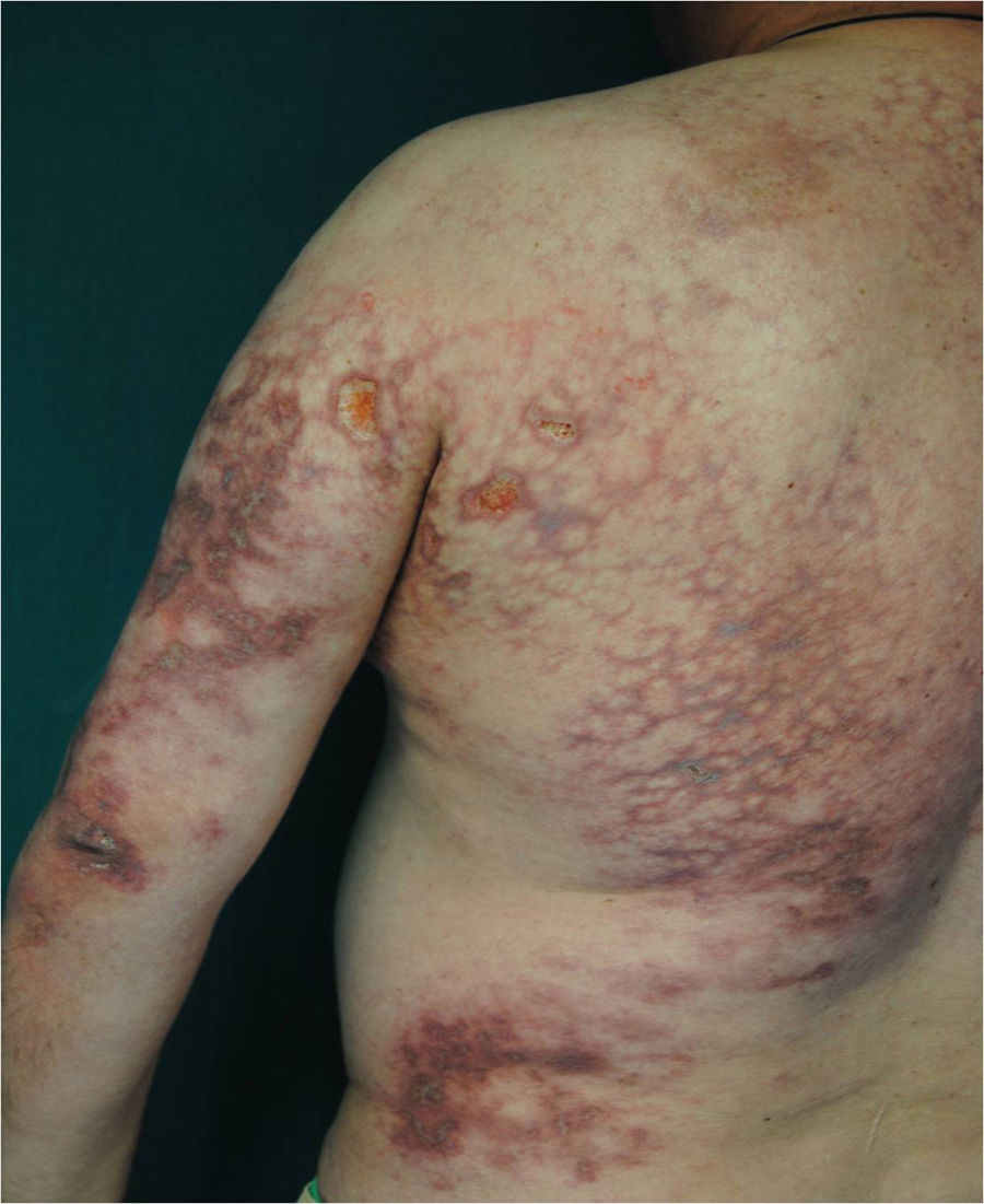
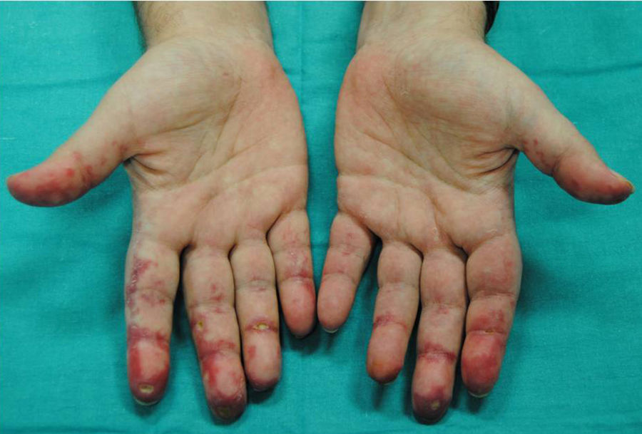
Our first patient was a 49-year-old man diagnosed with DM by cutaneous biopsy from the dorsal surface of the hand and the trunk (Fig. 1) in the context of edema, eyelid erythema, and arthritis. Laboratory findings included an erythrocyte sedimentation rate of 64 mm/h, a ferritin level of 609ng/mL, and aspartate transaminase and alanine transaminase levels both of 107U/L. On diagnosis of amyopathic DM, we started treatment with oral prednisone (1mg/kg/d) plus weekly doses of methotrexate (10mg). When painful ulcers developed on the palmar surfaces of the fingers (Fig. 2), plantar surfaces of the feet, and the trunk, bosentan (62.5mg/12h) and cyclophosphamide (1000mg/cycle) were started. After the second cycle the patient was admitted for sudden dyspnea and suspicion of interstitial pneumonia related to immunosuppression. A chest computed tomography scan showed dense subpleural reticulation in both lung fields. The patient's condition worsened in spite of antibiotic treatment and admission to the intensive care unit under mechanical ventilation. A rash developed and was biopsied. Histology showed extensive epidermal necrosis, leading to suspicion of toxic epidermal necrolysis. The patient was transferred to the burns unit of Hospital Universitario de la Paz. The skin condition resolved with administration of immunoglobulins, but the respiratory symptoms continued to worsen. Interstitial lung disease due to amyopathic DM was suspected. We started treatment with rituximab and plasmapheresis. A broader immunologic study (immunoblotting) confirmed the presence of anti-MDA-5 antibodies and the absence of transcriptional intermediary factor 1 autoantibodies (anti-TIF-1γ). The patient died from respiratory failure in spite of all measures.


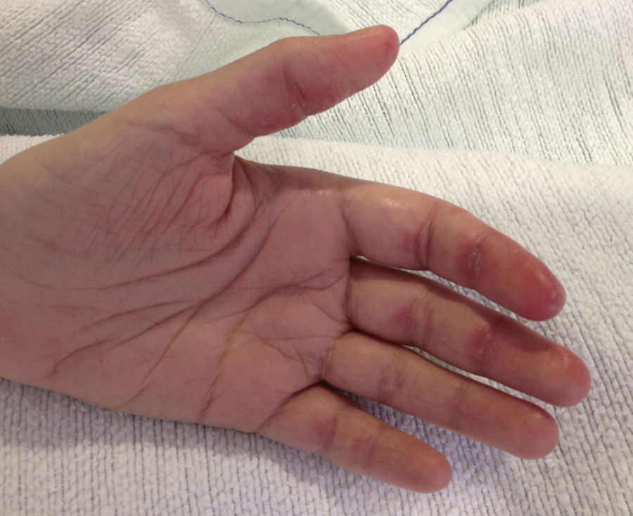
The second patient was a 31-year-old woman admitted with fever, dyspnea, and arthritic joint pain in her hands as well as lesions on her palms (Fig. 3) and elbows. An extensive test battery revealed vacuolar dermatitis at the dermal–epidermal junction, an aspartate transaminase level of 172U/L, an alanine transaminase level of 109U/L, elevated ferritin level of 1185ng/mL, an erythrocyte sedimentation rate of 64 mm/h, an anti-Sjögren's-syndrome-related antigen A antibody level of 103.80U/mL), and anti-MDA-5 positivity (by immunoblotting). Lung function tests showed restriction with a bilateral interstitial pattern in the lower lobes. Treatment with prednisone (1mg/kg/d), cyclophosphamide in bolus form, and hydroxychloroquine (200mg/d) was ordered. The patient did not improve on that regimen, so mycophenolate mofetil (2g/d) was substituted for cyclophosphamide. Slow improvement was noted, and the patient remained stable when the prednisone dosage was reduced to 10mg/d.

Anti-MDA-5 antibodies have been reported in up to 80% of amyopathic DM cases, and in 60% to 100% of cases of progressive interstitial lung disease in different series.3–5 The possible presence of skin signs has been noted in this clinical variant in recent years. Radiologic findings different from those usually described for DM have also been noted. Fiorentinoetal3 found that 10 in a series of 77 patients with DM showed anti-MDA-5 positivity and that this finding was significantly associated with hand edema, arthritis, skin ulcers, palmar macules and papules, mechanic's hands, alopecia, panniculitis, elbow erythema, and oral ulcers. However, Labrador-Horrilloetal6 were unable to confirm those findings in a Mediterranean series, possibly because both studies were retrospective and done in different populations. The discrepancies might also be due to small sample size, since this condition is uncommon and conclusions are difficult to draw. In our series of Caucasian patients, we saw several of the signs Fiorentinoetal reported (eg, arthritis, joint pain, hand edema, ulcers, and palmar papules). The lesions in our first patient (Fig. 1) were severe, and the maculopapular lesions both patients had between their fingers were very similar (Figs. 2 and 3). Our second patient's skin signs were milder, but we note that lesions as subtle as palmar macules were key to this diagnosis. The skin signs described by Fiorentinoetal have been reported in other cases along with skin ulcers, especially around the nail.3,7 Narangetal8 concluded that skin ulcers in DM might be related to anti-MDA-5 antibodies, predict lung involvement, and tend to be caused by vascular compromise. Finding these signs in the context of DM, therefore, can suggest a clinical strategy and call for anti-MDA-5 testing, which is not routinely ordered in most centers.
A noteworthy laboratory result is that the creatine kinase level tends to be normal whereas the components of liver function tests and ferritin tend to be very high,9 as we observed in our patients. High ferritin levels in DM suggest the likelihood of anti-MDA5 positivity and rapid progression of lung disease.
Radiologic findings also vary. The most common pattern in anti-MDA-5–positive cases is a subpleural ground-glass opacity in lower lung fields.10
In summary, we have described 2 cases of anti-MDA-5 positivity in 2 patients with amyopathic DM and characteristic skin signs such as ulcerations around the nails and palmar papules. We have attempted to describe this rare condition, which has certain clinical features that differ from classical DM. Understanding this phenotype will contribute to improved diagnosis and better follow-up of patients with a condition that requires us to watch for possible lung involvement.
Conflicts of InterestThe authors declare that they have no conflicts of interest.
Please cite this article as: Barrientos N, Sicilia JJ, Vega MJMd, Dominguez JD. Dermatomiositis anti-MDA-5 positivas. Descripción de clínica cutánea y sistémica a propósito de dos casos. Actas Dermosifiliogr. 2018;109:188–190.


